Tour
1: Basics
Designed for
beginners
This chapter should help you to go familiarly with the Pannoramic
scanners P250, SCAN 150,
Overview
of Pannoramic scanners
Use Microscope or Scanner instead?
In our descriptions, the term “microscope” and “scanner” is often used
likewise. All of our microscopes are scanners also and all of our scanners
include magnifying of the tissue like a microscope does. Often the relevant
expression is used to focusing the attention to the more important task at the
appropriate moment, but this is not done consequently. So we can assume, the
used term “microscope” or “scanner” has no important difference in our aspects.
What is a scanner?
 The term “scanner” used by 3DHISTECH includes a
microscope unit to magnifying the object (tissue) to be scanned. The tissue
itself is scanned FOV by FOV (FOV means field of view; the area of the tissue,
captured by the scan camera with 1exposure) and then, these fields are assembled
by the software to a virtual slide (or virtual tissue).
The term “scanner” used by 3DHISTECH includes a
microscope unit to magnifying the object (tissue) to be scanned. The tissue
itself is scanned FOV by FOV (FOV means field of view; the area of the tissue,
captured by the scan camera with 1exposure) and then, these fields are assembled
by the software to a virtual slide (or virtual tissue).
To reach this, because each virtual tissue may be assembled from more
100 FOVs, the tissue must be moved in X- and Y-direction in relation to the
objective pupil. So we can say, the X-Y-stage unit, equipped with the specimen
holder is a part of the scanner.
In opposite to this, the focus unit (contains the objective, the
condenser and the focusing mechanics) is a part of the microscope unit; a part
of the optical path.
Both units are mounted together on the scanner plate and the assembled
scanner plate makes the scanner unit.
This example shows that the term is floating or, the boarder between
scanner and microscope can often not be defined exactly.
Watch video: Scan a tissue_P250
A short history
 The development of
the first scanner started in the early 2000’s years.
The development of
the first scanner started in the early 2000’s years.
The goal should be to create a microscope that is able to scan glass slides
with tissues automatically; to creating a virtual tissue and this virtual
tissue (or virtual slide) should be stored on a HDD.
Furthermore, the removal of the finished slide and the insertion of the
next slide to be scanned should be done automatically, without interaction of
the user.
Allowed slide dimensions
was defined as
Length: 75.00 to 76.00
mm
Width: 25.00 to 26.00
mm
Thickness: 00.95 to 01.05
mm
After some development and test versions, the scanner MIRAX SCAN was born in the year 2004.
![]() DIGITAL MICROSCOPY and
DIGITAL PATHOLOGY
DIGITAL MICROSCOPY and
DIGITAL PATHOLOGY
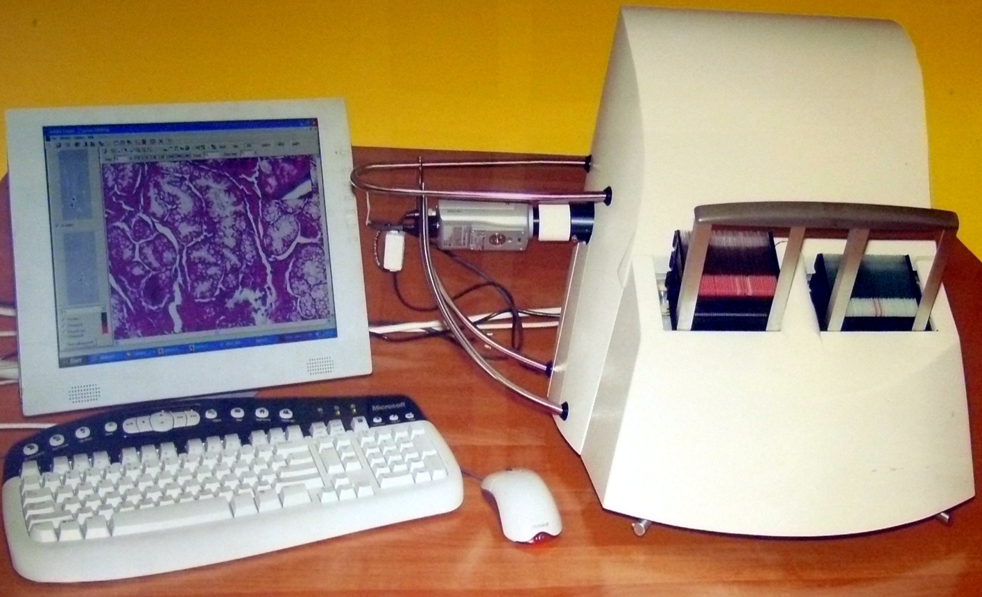 If the scan
procedure of a slide is finished, the virtual slide (the result of the scan process)
can be made visible on the computer screen with the viewer program.
If the scan
procedure of a slide is finished, the virtual slide (the result of the scan process)
can be made visible on the computer screen with the viewer program.
The viewer program with its various features and options is used to
analyze, qualify and to prepare the virtual tissue with annotations regarding
interesting or abnormal passages in the tissue.
Pannoramic viewer options and
features; today
· 3D
· OnlineTeleconsultation
· TMA
· FLViewer
· BookmarkExport
· IHLab the
new name is “TumorBoard”
· HistoQuant
· DataVisualisation
· MarkerCounter
· CytoFISHQuant
· NuclearQuant
· MembraneQuant
· TumorBoard
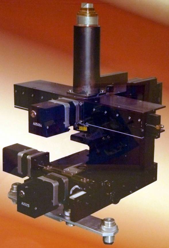
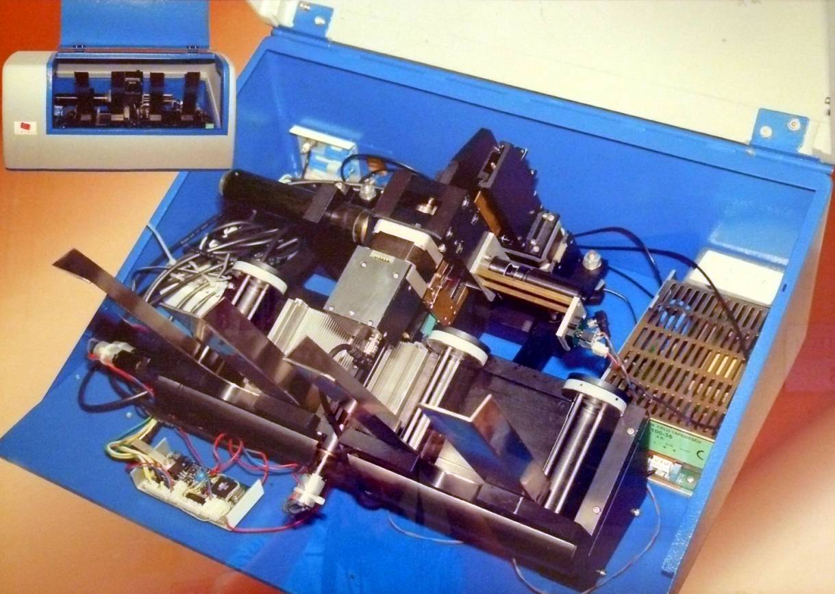
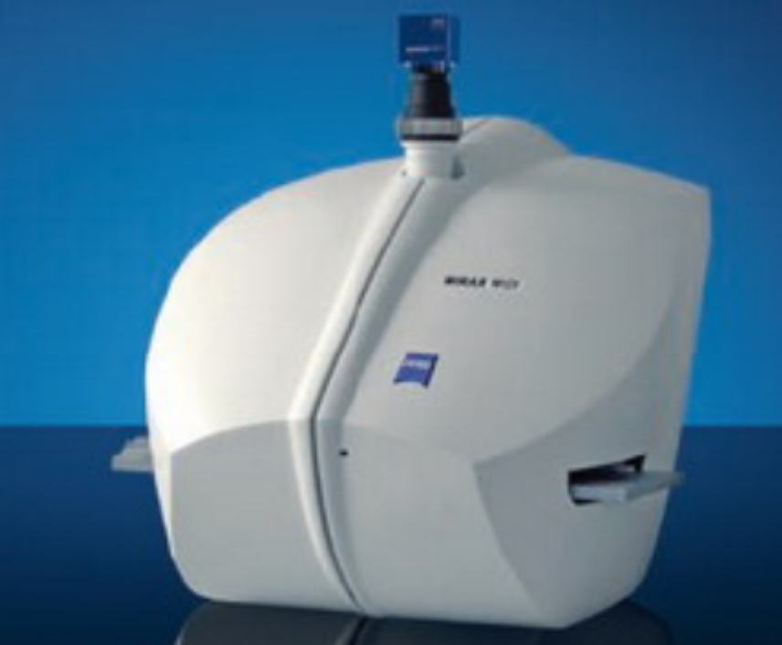
MIRAX SCAN
 This scanner (distributed
and serviced by Carl Zeiss G.m.b.H) uses magazines with 50 slide positions, and
6 magazines could be loaded in the input stack. With this solution, 300 slides
could be scanned during 1 scan session.
This scanner (distributed
and serviced by Carl Zeiss G.m.b.H) uses magazines with 50 slide positions, and
6 magazines could be loaded in the input stack. With this solution, 300 slides
could be scanned during 1 scan session.
· Since 2004 on the
market
· The slides are hold
in magazines with 50 slide positions
· 6 magazines can be
hold in the input / output stack during 1 session.
· Designed for
brightfield an fluorescent scan sessions.
· The scan speed
depends highly on the used computer configuration and the speed of the camera.
Allowed slide dimensions are defined as
Length: 75.00 to 76.00
mm
Width: 25.00 to 26.00
mm
Thickness: 00.95 to 01.05
mm
The SCAN exists in a full version (SCAN_FL) where the Reflector turret unit
is included and a more cost-effective version (SCAN _BF) without the Reflector
turret unit, so only brightfield scan sessions are possible.
Today, these machines are almost all upgraded to the version SCAN 150.
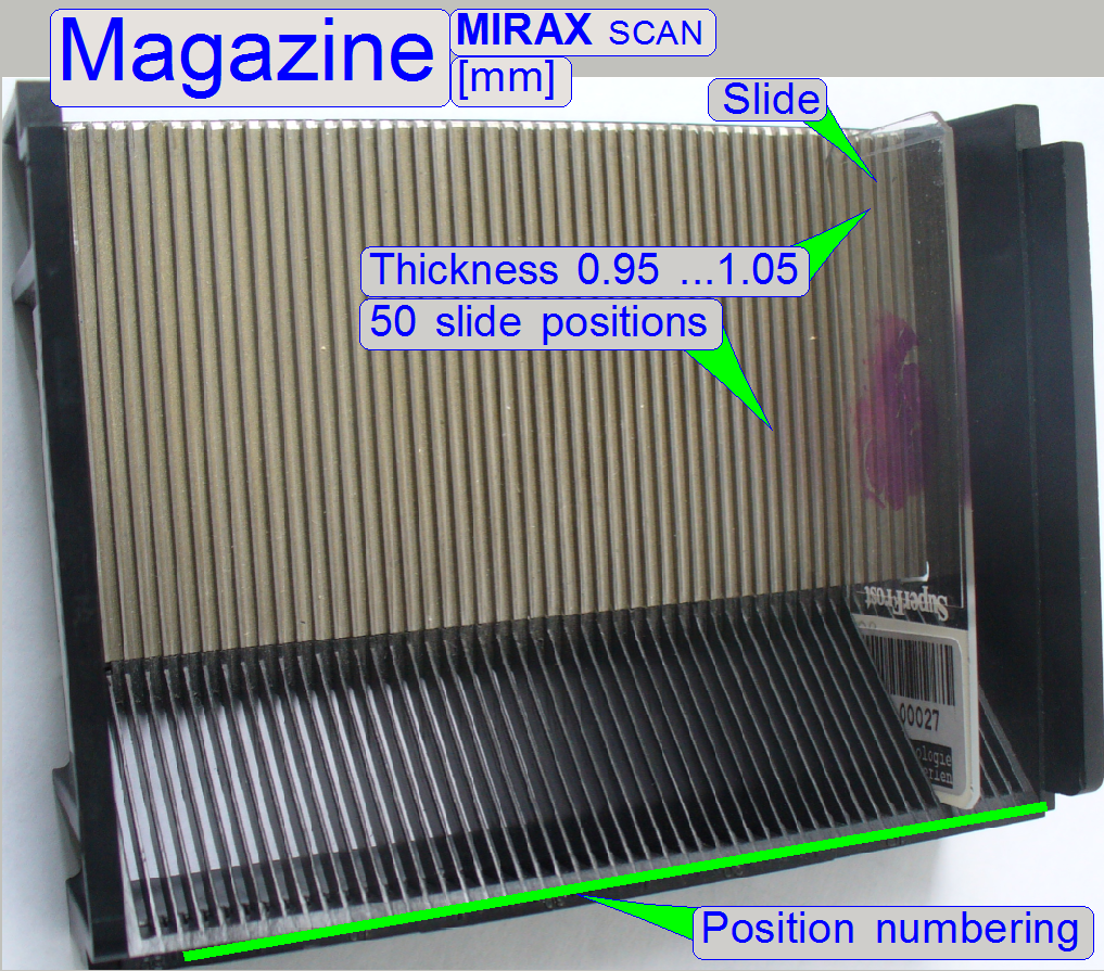
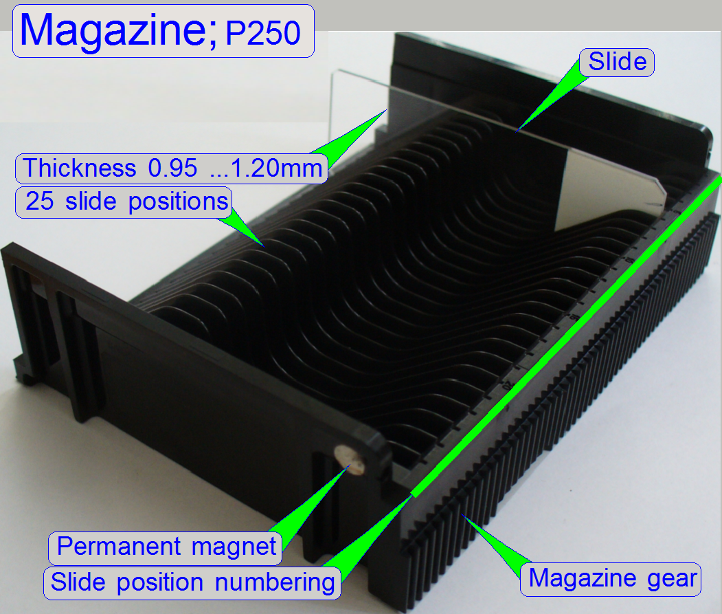
The magazines used in the scanners “MIRAX SCAN”, before upgrading to SCAN 150
· The
magazine contains 50 slide positions
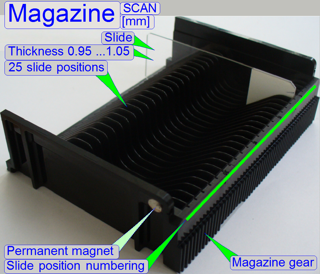 The external
dimensions of the magazines are unchanged; only the construction of the slide
positions was modified and changed to the half.
The external
dimensions of the magazines are unchanged; only the construction of the slide
positions was modified and changed to the half.
“SCAN
· To reduce the
amount of different magazines, all the magazines with 25 slide positions has a
permanent magnet implemented, but this magnet acts only in the P250.
SCAN 150
To improve the slide loading stability of the MIRAX SCAN the magazine and the slide loading mechanics was
modified for the use of magazines with only 25 slide positions. This way, the
stability of the slide insert and slide removal procedure could be improved.
· Since the end of
the year 2008 on the market; almost all MIRAX SCAN scanners are upgraded to
this version.
· The slides are
hold in magazines with 25 slide positions; the useable slide dimensions are
unchanged.
· 6 magazines can be
hold in the input and output stack during 1 session.
· Designed for
brightfield and fluorescent scan sessions.
· The scan speed
depends highly on the used computer configuration and the speed of the camera.
Allowed slide dimensions
are defined as
Length: 75.00 to 76.00
mm
Width: 25.00 to 26.00
mm
Thickness: 00.95 to 01.05
mm
The SCAN 150 exists in a full version (SCAN_FL) where the Reflector
turret unit is included and a more cost-effective version (SCAN _BF) without
the Reflector turret unit, so only brightfield scan sessions are possible.
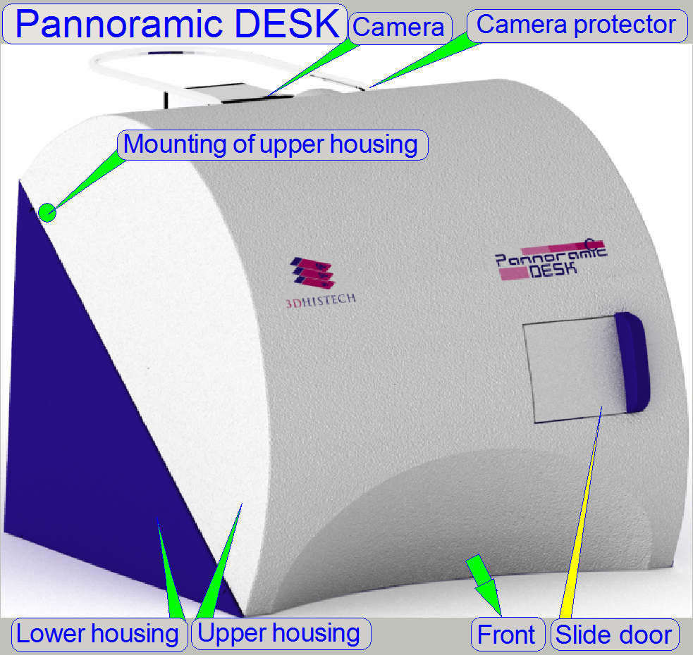
MIRAX DESK
 For small
pathology laboratories the MIRAX DESK version was created. This scanner
contains in practice a scanner unit only and the slides are inserted or
exchanged manually; the fluorescent illumination unit (Reflector turret unit)
is left out.
For small
pathology laboratories the MIRAX DESK version was created. This scanner
contains in practice a scanner unit only and the slides are inserted or
exchanged manually; the fluorescent illumination unit (Reflector turret unit)
is left out.
· Since 2006 on the
market
· The slides are
inserted or removed manually via the slide door.
· Used in small
laboratories with a small amount of slides to be scanned.
· Designed for brightfield
scan sessions only.
· The scan speed
depends highly on the used computer configuration and the speed of the camera.
Allowed slide dimensions are defined as
Length: 75.00 to 76.00
mm
Width: 25.00 to 26.00
mm
Thickness: 00.95 to 01.05
mm
MIRAX
The two versions, MIRAX SCAN
and MIRAX DESK are used in very big
and very small pathology laboratories. The users asked for a machine that can
be used in middle large laboratories, where the manual handling of slides is
too slow and the capacity of the MIRAX
SCAN is not efficiently used.
These requirements resulted in the MIRAX
· Since 2007 on the
market
· The slides are
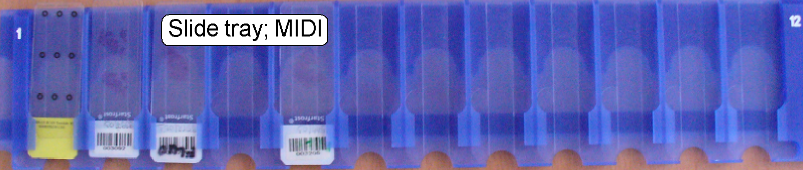
inserted / removed automatically, via a slide holder tray.
· 12 slide bays in
the slide holder tray allowing an automatic scan session of about ½ to
¾ hour, depending on the tissue size.
· Used in
laboratories with middle large amount of slides to be scanned.
· Designed for
brightfield scan sessions and fluorescent scan sessions likewise.
· The scan speed
depends highly on the used computer configuration and the speed of the camera.
Allowed slide dimensions
are defined as
Length: 75.00 to 76.00
mm
Width: 25.00 to 26.00
mm
Thickness: 00.95 to 01.05
mm
The
Slide tray of the
At the end of the year 2009 the contract with Carl Zeiss G.m.b.H was
finished. Because MIRAX™ is a trademark of Carl Zeiss G.m.b.H, the name
“Pannoramic” (a combination of
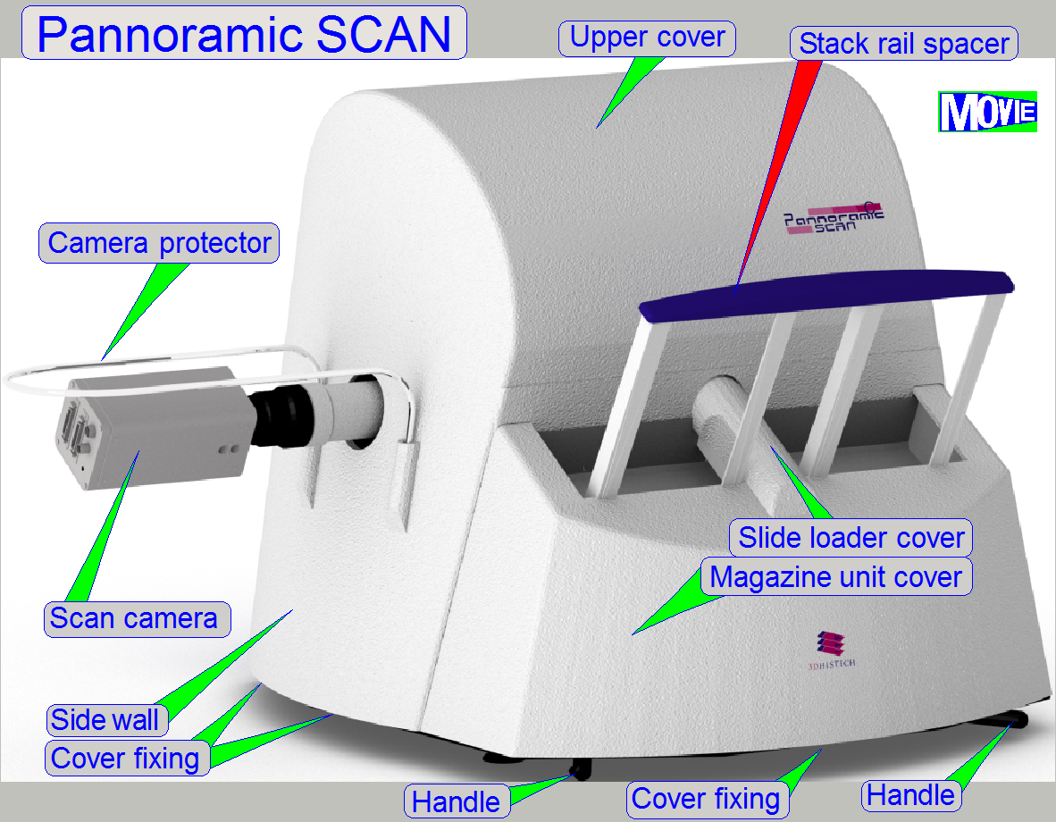
In this time, the name of the scanners changed to Pannoramic SCAN,
Pannoramic MIDI and Pannoramic DESK and 3DHISTECH contracted distributors world
wide for distribution and services of Pannoramic scanners and 3DHISTECH
products.

· The slides are
hold in magazines with 25 slide positions
· 6 magazines can be
hold in the input / output stack during 1 session.
· Designed for
brightfield an fluorescent scan sessions.
· The scan speed
depends highly on the used computer configuration and the speed of the camera.
· Today, all
references to the SCAN, PSCAN or SCAN150 referring to this version of scanner.
Allowed slide dimensions are defined as
Length: 75.00 to 76.00
mm
Width: 25.00 to 26.00
mm
Thickness: 00.95 to 01.05
mm
The SCAN exists in a full version (SCAN_FL) where the Reflector turret unit
is included and a more cost-effective version (SCAN _BF) without the Reflector
turret unit, so only brightfield scan sessions are possible.


· The slides are
inserted / removed automatically, via a slide holder tray.
· 12 slide boxes in
the slide holder tray
· Used in
laboratories with middle large amount of slides to be scanned.
· Designed for
brightfield scan sessions and fluorescent scan sessions likewise.
· The scan speed
depends highly on the used computer configuration and the speed of the camera.
Allowed slide dimensions are defined as
Length: 75.00 to 76.00
mm
Width: 25.00 to 26.00
mm
Thickness: 00.95 to 01.05
mm
The

· The slides are
inserted / removed manually via the slide door.
· Used in small
laboratories with a small amount of slides to be scanned.
· Designed for
brightfield scan sessions only.
· The scan speed
depends highly on the used computer configuration and the speed of the camera.
Allowed slide dimensions
are defined as
Length: 75.00 to 76.00
mm
Width: 25.00 to 26.00
mm
Thickness: 00.95 to 01.05
mm

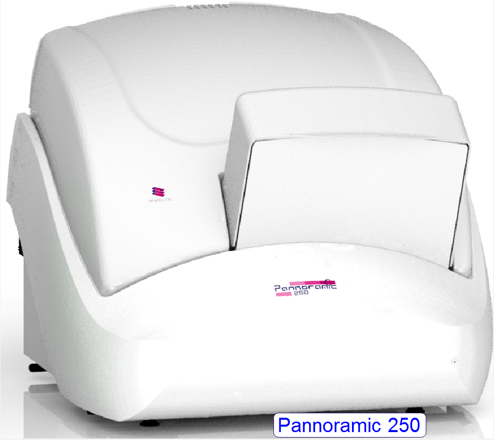 The Pannoramic 250
(P250) includes all possibilities and features like different camera selection
for fluorescent scan sessions and brightfield scan sessions software controlled,
selection of the objective 20x or 40x magnification for brightfield and
fluorescent scan sessions likewise.
The Pannoramic 250
(P250) includes all possibilities and features like different camera selection
for fluorescent scan sessions and brightfield scan sessions software controlled,
selection of the objective 20x or 40x magnification for brightfield and
fluorescent scan sessions likewise.
· The possible slide
thickness was extended to be 1.20mm
maximal
Allowed slide dimensions are defined as:
Length: 75.00 to 76.00 mm
Width: 25.00 to 26.00 mm
Thickness: 00.95 to 01.20 mm
· Since 2011 on the
market
· The slides are
hold in magazines with 25 slide positions
· 9 magazines can be
hold in the input / output stack and 1 magazine in the feeder channel, so the
amount of slides in 10 magazines can be scanned during 1 session.
· The specimen
holder is modified; so slides with a thickness of 1.20mm can be inserted.
· Flashlight BF
illumination and the use of higher speed cameras allows higher scan speeds.
· Designed for
brightfield an fluorescent scan sessions.
· Exchange of the
objective 20x or 40x magnification; software controlled between 2 slide scan
sessions.
· Exchange of the
camera, software controlled, between BF and FL slide scan sessions.
The P250 exists in a full version (P250_FL) where the Reflector turret
unit is included and a more cost-effective version (P250_BF) without the
Reflector turret unit, so only brightfield scan sessions are possible.
· In
opposite to the SCAN, the presence of the permanent magnet is now required.
The permanent magnet signals in the magazine input stack the presence of
a magazine while in the output stack the state “output stack full” is so detected.

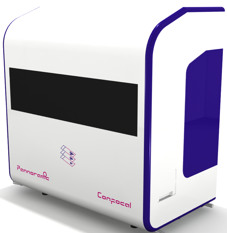 The Pannoramic
Confocal (PCON) scanner gives the possibility to scan slides in confocal mode.
The Pannoramic
Confocal (PCON) scanner gives the possibility to scan slides in confocal mode.
The scanner is based on a modular
Since 2014 on the market
· The slides are
inserted and removed automatically via a slide holder tray.
· 12 slide bays in
the slide holder tray allowing a more or less autonomic work of the scanner.
· Used in
laboratories with middle large amount of slides to be scanned and for research
and development purposes in pathology, pharmacy and biology.
· Designed for
brightfield scan sessions, fluorescent and con-focal fluorescent scan sessions.
Allowed slide dimensions are defined as
Length: 75.00 to 76.00 mm
Width: 25.00 to 26.00 mm
Thickness: 00.95 to 01.05 mm
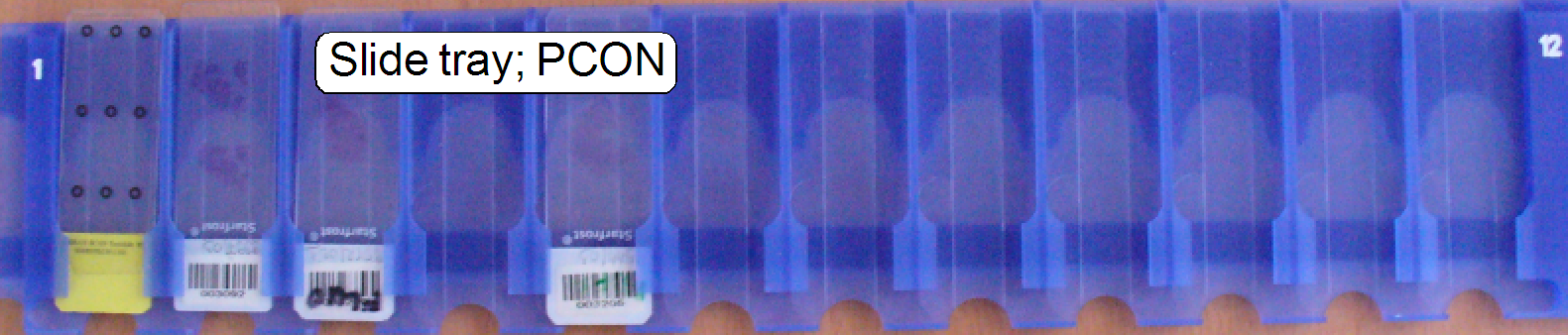 Slide
holder tray of the PCON; it allows an automatic scan session of about
¾ to 1 hour, the scan time of the entirely filled tray depends highly on
the tissue size .
Slide
holder tray of the PCON; it allows an automatic scan session of about
¾ to 1 hour, the scan time of the entirely filled tray depends highly on
the tissue size .
Optics and illumination
For beginners (as well as for experienced technicians) it is very
important to know the influence of optical means like lenses, mirrors and
filters on light rays; the most efficient illumination of the tissue and other
techniques in microscopy.
A lot of excellent prepared explanations about optics, illumination,
microscopy, brightfield illuminated and fluorescent excited scan methods and
others can be found in the internet.
The second main part is fine mechanics, stepper motors and step control
of stepper motors.
The following chapters are highly
recommended:
Microscopy from the
beginning; PDF-file;
by © Carl Zeiss GmbH;
Oberkochen,
Ernst Abbe und das Mikroskop YouTube German
language
Bildentstehung bei der Sammellinse © Dr. Andreas Rueff more chapters, German; Schrittweise,
graphische Erläuterung der Bildentstehung bei Verwendung von Linsen
Picturing by converging lens YouTube with English subtitles;
explanations are also visualized graphically in steps
Image
Formation with Converging Lenses Interactive
Java tutorial
Education in Microscopy
and Digital Imaging © the Carl Zeiss Microscopy
Online Campus
Köhler
illumination; © Wikipedia
Physics of
light and color; ©
2012 Olympus America Inc.; all rights reserved.
Brightfield
microscopy; ©
2012 Olympus America Inc.; all rights reserved.
Fluorescence
microscopy; ©
2012 Olympus America Inc.; all rights reserved.
Other chapters related to
optics and microscopes are also very interesting and the interactive tutorials
are very helpful to understand the behavior of the light rays.
Bayer mosaic and
“Debayering” see: “Bayer filter” Wikipedia
“Color processing with Bayer Mosaic
sensors” stored in this description Copyright
© MATRIX
Vision GmbH
“Farbverarbeitung mit Bayer-Mosaic
Sensor” stored in this description Copyright © MATRIX
Vision GmbH
“Color filter array” Wikipedia
“Demosaicing” Wikipedia
“Bayer-Sensor” Wikipedia de
FOV
and Preview Area “Theory”; “Sample scan
process”; “Optical path and Field Of View”; “Influence of the camera
adapter”
Precautions Important precautions related to
components, units and Pannoramic scanners
Stepper or step motors
In Pannoramic scanners the stepper motors are always driven in micro
stepping mode; so please focus your attention on it.
The following chapters are highly recommended:
Stepper motors Wikipedia
Stepper
motors © Nanotec the
interactive, animated demonstration shows how stepper motors working.
About
basics, theory and principles please refer to:
http://www.solarbotics.net/library/pdflib/pdf/motorbas.pdf
Stepper
motor basics (stored)
Drive circuit basics (stored)
Stepper motor and driver (stored)
External
recirculation diodes (stored)
Stepper motor
driving (stored)
Stepper motors 2011 (stored)
Power and control (P250) “Stepper
motor”
End; you may continue with step 2, “Optics in the SCAN, MIDI and DESK”