This case study explores how 3DHISTECH’s Digital Pathology and Tissue Microarray Solutions – including the Pannoramic Confocal, Scan II, MIDI III, 150, 480, 1000 digital scanners, TMA molmed, TMA Master II, and software solutions such as QuantCenter, SlideManager, and WebViewer – address key challenges in toxicology research.
Key Challenges in Toxicology Research
High Variability in Tissue Analysis
Manual slide examination can lead to inconsistencies in toxicological findings due to subjective interpretations
Time-Intensive Histopathology Workflows
Traditional toxicology studies require extensive manual slide preparation, staining, and evaluation, slowing down drug development
Limited Throughput for Large-Scale Toxicity Screening
Toxicology often requires testing multiple compounds across thousands of slides, which is difficult to achieve with conventional microscopy
Difficulty in Biomarker Quantification for Drug-Induced Tissue Damage
Assessing cellular responses to toxins requires precise biomarker quantification, which is challenging with manual methods
Lack of Standardized Sample Handling and Centralized Data Management
Maintaining consistency in sample preparation, imaging, and data processing is crucial for regulatory compliance
№1. Digital Pathology Scanners for High-Throughput and Precision
Pannoramic Confocal Digital Scanner

Key Features
- Confocal Z-Stack scanning for 3D imaging.
- Advanced fluorescence capabilities for biomarker detection.
- Ideal for deep tissue analysis and toxicological screening.
Pannoramic Scan II Digital Scanner

Key Features
- 150-slide capacity with brightfield & fluorescence imaging.
- High-throughput tissue screening for drug safety testing.
Pannoramic MIDI III Digital Scanner
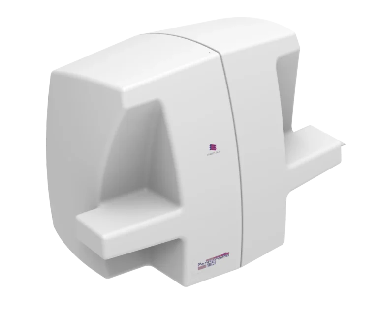
Key Features
- 12-slide capacity with AI-driven analysis.
- Precise biomarker quantification and detailed morphology studies.
Pannoramic 150 Digital Scanner
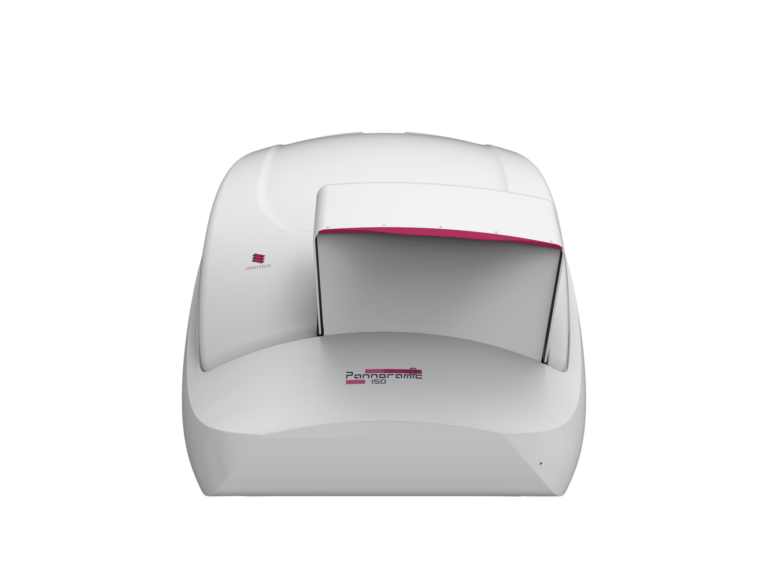
Key Features
- 150-slide capacity with 10x fluorescence objective.
- Faster fluorescence analysis for toxicity detection.
Pannoramic 480 Digital Scanner
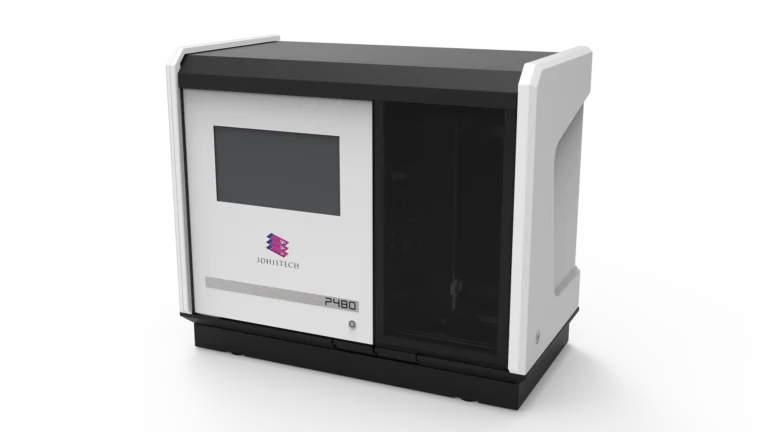
Key Features
- 480-slide capacity with polarization technology.
- Best for large-scale toxicology studies.
Pannoramic 1000 Digital Scanner
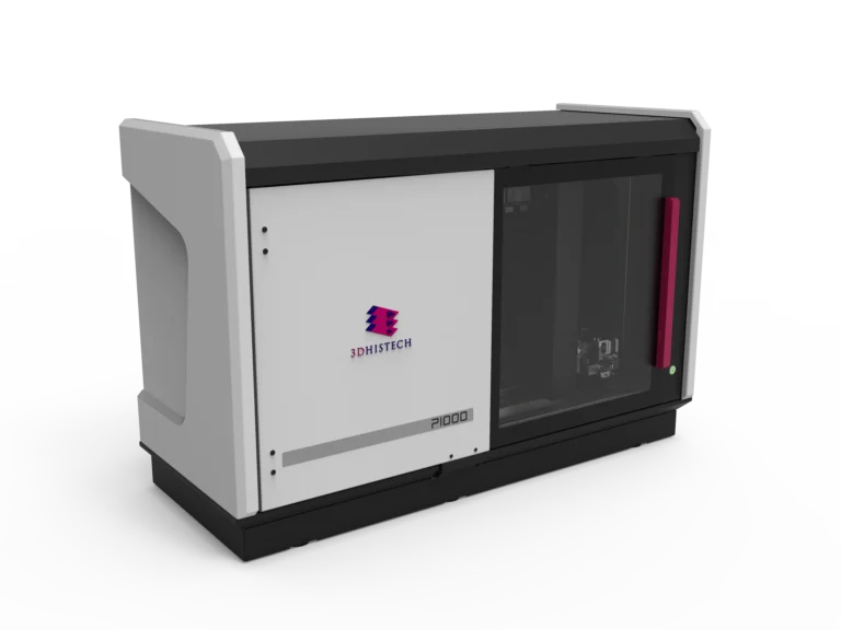
Key Features
- 1200-slide capacity with a water immersion system.
- Designed for ultra-high-volume toxicology research in pharmaceuticals.
№2. Tissue Microarray (TMA) Solutions for High-Throughput Toxicology
TMA molmed

Key Features
- High-throughput microarray processing with 558 cores per block.
- Molecular biomarker quantification using PCR, dPCR, and sequencing workflows.
- Enables batch comparison of drug-treated vs. control samples.
TMA Master II

Key Features
- Compact, fully automated system for standardized tissue sampling.
- Can create four identical TMA blocks, ensuring reproducibility.
- Integrated barcode tracking & digital slide overlay for sample validation.
№3. Advanced Software Solutions for Toxicology
SlideViewer & WebViewer
Remote viewing and annotation, facilitating multi-site collaboration in toxicology research
Implementation
Implementation and Results
Scenario 1: Pharmaceutical Company Conducting Preclinical Drug Toxicity Studies
Problem
- A leading pharmaceutical company needed to test hepatic toxicity of a new drug.
- The study required analyzing 1,000+ liver tissue samples for biomarkers indicating cellular damage.
Implementation
- Pannoramic 1000 and 480 scanners digitized all liver tissue samples for AI-powered image analysis.
- TMA molmed extracted core tissue samples for biomarker validation.
- QuantCenter’s HistoQuant module quantified liver cell necrosis and inflammation.
- WebViewer enabled remote validation by pathologists.
Results & Impact
- 80% reduction in analysis time through automated biomarker quantification.
- 40% reduction in reagent costs by consolidating samples into TMAs.
- 30% faster preclinical testing, accelerating drug approval.
Scenario 2: Toxicological Screening of Environmental Pollutants
Problem
A research institute studying air pollution’s impact on lung tissues needed high-resolution imaging for toxicological assessments.
Implementation
- Pannoramic Confocal provided 3D lung tissue imaging to detect morphological changes.
- TMA Master II created standardized tissue microarrays for multi-year tracking of toxicity effects.
- QuantCenter’s PatternQuant module identified cellular alterations linked to pollution exposure.
Results & Impact
- Deeper insights into structural damage using 3D tissue imaging.
- Automated biomarker analysis enabled long-term studies.
- Facilitated collaboration between toxicologists and environmental scientists, leading to breakthrough publications.
Scenario 3: CRO Enhancing Efficiency in Toxicology Testing
Problem
A CRO needed faster, high-throughput toxicology testing for multiple clients.
Implementation
- Pannoramic Scan II & MIDI III provided fast, high-resolution slide scanning.
- TMA molmed & TMA Master II optimized sample preparation.
- AI-driven QuantCenter modules automated toxicity scoring.
Results & Impact
- 50% increase in study throughput for CROs, allowing them to take on more clients.
- Improved accuracy and consistency in toxicity scoring.
- Faster turnaround times, increasing client satisfaction and revenue.
Who Benefits from These Solutions?
Pharmaceutical & Biotech Companies
• Faster, cost-effective drug safety screening.
• Higher reproducibility in preclinical studies.
Toxicology Research Labs
- Standardized biomarker quantification across multiple test groups.
- Improved tissue damage assessment in drug trials.
Contract Research Organizations (CROs)
• Ability to process higher sample volumes with greater efficiency.
• Reduces workload for toxicopathologists.
Regulatory Agencies (FDA, EMA, etc.)
- Standardized workflows improve transparency and traceability.
- Faster review of drug safety data.
Remodeling Toxicology with Digital Pathology & TMAs
The integration of Whole Slide Imaging (WSI) and Tissue Microarray (TMA) solutions significantly enhances high-throughput toxicology workflows. These solutions enable:
- Rapid processing of hundreds of tissue samples
- Standardized biomarker quantification
- More efficient drug-induced tissue damage analysis
- Regulatory-compliant data management
By combining TMAs with Whole Slide Imaging (WSI) and AI-powered analysis, toxicology research becomes faster, more reproducible, and cost-effective, ultimately accelerating drug safety assessments and environmental toxicology studies.