To address these limitations, digital pathology and tissue microarray (TMA) solutions provide high-resolution imaging, automated analysis, and scalable data management for pharmacological applications. This case study explores how whole slide imaging (WSI), AI-powered quantification, and automated tissue microarray solutions enhance drug efficacy testing, biomarker validation, and toxicology studies.
Key Challenges in Pharmacology Research
High-Throughput Screening for Drug Discovery
• Pharmacology research requires large-scale tissue imaging and analysis to evaluate drug efficacy and toxicity.
• Manual slide scanning and microscopic analysis are slow and inefficient for preclinical studies.
Multiplex Imaging for Drug Localization and Biomarker Studies
• Understanding drug-tissue interactions requires fluorescence multiplexing and 3D imaging.
• Conventional imaging techniques often fail to capture subcellular structures and molecular distributions.
AI-Powered Quantification of Drug Effects
• Assessing biomarker expression, cellular morphology, and molecular changes demands precise, automated analysis.
• Traditional histopathological evaluation is subject to human error and interobserver variability.
Tissue Microarray Integration for Large-Scale Biomarker Research
• Drug response studies require batch processing of tissue samples for comparative biomarker analysis.
• Manually constructing TMAs is slow and prone to errors, limiting the reproducibility of results.
Digital Data Management & Remote Collaboration
- Large-scale pharmacology studies generate massive datasets requiring secure, centralized storage and global access.
- Research teams need real-time collaboration for multi-center clinical trials and translational research.
Optimizing Pharmacology
Solutions
№1. High-Throughput Drug Screening & Toxicology Studies
Key Benefits
- Automated scanning accelerates drug screening workflows.
- AI-driven image analysis detects drug-induced morphological changes.
- Z-stack imaging improves visualization of complex tissue structures.
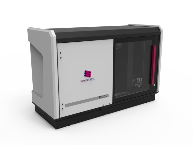
Pannoramic 1000 Digital Scanner
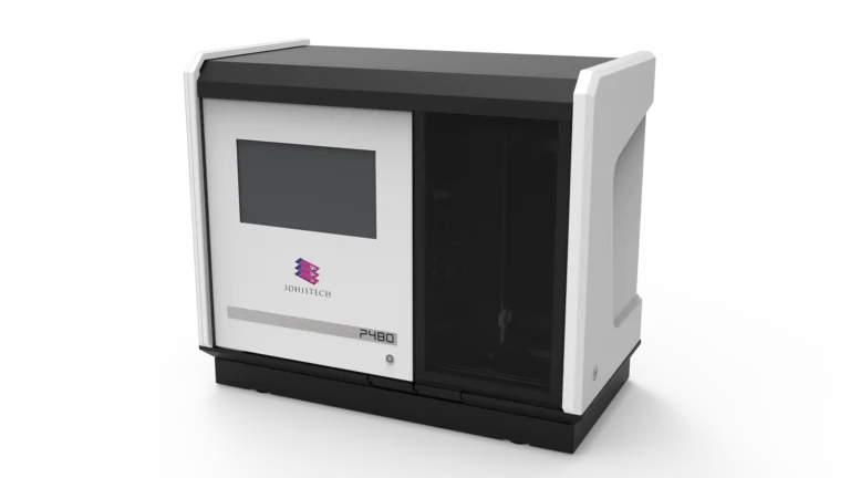
Pannoramic 480 Digital Scanner
Real-World Application
A pharmaceutical R&D lab tested a novel anticancer compound using Pannoramic 1000. High-throughput scanning reduced slide processing time by 60%, allowing for rapid preclinical toxicity assessment.
№2. High-Resolution Fluorescence Imaging for Drug Localization
Key Features
Key Benefits
- Enables precise tracking of drug distribution within tissues.
- High-resolution imaging supports fluorescence-based receptor-targeted pharmacology.
- Reduces photo-bleaching, enhancing fluorescence signal integrity.
Pannoramic Confocal Digital Scanner

Real-World Application
A neuroscience research center used Pannoramic Confocal to study neuroactive drug diffusion across the blood-brain barrier. 3D imaging provided subcellular-level insights into drug penetration dynamics.
№3. AI-Driven Biomarker Quantification & Digital Image Analysis
Key Features
Key Benefits
- Reduces interobserver variability in biomarker quantification.
- Enables rapid and reproducible tissue-based pharmacokinetics analysis.
- Supports quantitative drug efficacy assessments.
Software Solutions
Real-World Application
A biotech company used QuantCenter to analyze PD-L1 biomarker expression in immunotherapy-treated tumors. AI-driven quantification reduced analysis time from 3 days to a few hours, improving data consistency and decision-making in clinical trials.
№4. Tissue Microarray Solutions for Large-Scale Biomarker Studies
Key Features
Key Benefits
- Processes up to 250 cores per hour, accelerating batch testing.
- Improves standardization in large-scale biomarker research.
- Enables high-throughput drug screening with minimal sample usage.
TMA Grand Master

TMA molmed

Real-World Application
A cancer research institute studying chemotherapy-induced toxicity used TMA molmed to batch process 500 tissue samples. Automated core extraction and digital integration reduced experiment time by 75%, enabling faster biomarker validation.
№5. Digital Data Management & Remote Collaboration
Key Features
Key Benefits
- Secure, scalable storage for pharmacology datasets.
- Facilitates multicenter collaboration in drug trials.
- Enables cloud-based slide sharing for remote pathology analysis.
Software Solutions
Real-World Application
A global pharmaceutical company conducting a cardiovascular drug trial used SlideManager to coordinate tissue analysis across five research centers. Remote slide access accelerated data review and regulatory approvals.
Implementation, Impact & Results
High-throughput drug screening Pannoramic 1000 & 480 60% reduction in screening time, enabling rapid drug discovery.
Multiplex fluorescence imaging Pannoramic Confocal Improved drug localization studies & enhanced biomarker visualization.
AI-driven biomarker quantification QuantCenter Automated analysis reduced variability, saving 3 days per dataset.
Large-scale biomarker studies TMA molmed 4x faster tissue microarray processing, enhancing pharmacogenomics research.
Digital data management & collaboration SlideManager & WebViewer Enabled global research teams to collaborate in real-time.
Who Benefits from These Solutions?
Pharmaceutical Companies
Faster drug development, scalable biomarker analysis, and AI-powered pathology.
Research Laboratories
High-throughput digital workflows, improved reproducibility, and faster data analysis.
Clinical Trial Teams
Secure, remote access to digital slides, accelerating biomarker validation and regulatory approvals.
Toxicology & Pharmacokinetics Researchers
Automated toxicity assessments, high-resolution imaging for dose-response studies.
The integration of digital pathology and tissue microarray technologies transforms pharmacology research, enabling faster, more reproducible, and scalable solutions for drug discovery, biomarker validation, and toxicology studies.
By automating high-throughput imaging, AI-powered analysis, and digital collaboration, these solutions provide pharmaceutical companies, research institutions, and clinical trial teams with unparalleled efficiency, accuracy, and scalability.