By digitizing biopsies, histological samples, and cytopathology specimens at ultra-high resolution, these solutions enable early cancer detection, streamlined workflow automation, and enhanced remote collaboration, transforming oncology diagnostics.
Key Challenges in Cancer Diagnostics
High Case Volumes & Growing Demand
Pathologists face increasing workloads, requiring faster and more efficient digital workflows.
Need for High-Resolution Imaging
Accurate cancer detection depends on identifying subtle morphological variations in tissue samples.
Complexity of Cancer Subtypes
Precise imaging and quantification are needed for different cancer types, from solid tumors to hematological malignancies.
Delayed Diagnoses Due to Slide Transport
Physical slide shipping for second opinions and consultations can slow down critical patient management.
Data Management & Integration Issues
Pathology labs need seamless LIS/HIS compatibility to ensure accurate case tracking and reporting.
The Pannoramic DX Whole Slide Imaging Solutions optimize cancer diagnostics by offering:
Ultra-High-Resolution Imaging
Capture intricate cellular details at up to 100x magnification (0.1 μm/pixel resolution).
Automated Tissue Detection & AI-Powered Workflows
Enhance accuracy and minimize manual effort.
Multilayer Z-Stack & Extended Focus Imaging
Ideal for complex oncology cases requiring multiple focal planes.
Telepathology for Remote Collaboration
Enable pathologists to consult specialists worldwide in real time.
Seamless LIS/HIS Integration
Ensure smooth data flow and case management.
Optimizing Oncology Pathology for Cancer Diagnostics
Pannoramic DX Digital Solutions for Oncology Pathology
№1. Best for large hospitals, high-throughput diagnostic labs, and cancer research centers.
Pannoramic 250 FLASH III Digital Scanner

Key Features
- Capacity: Scans up to 300 slides for uninterrupted workflow.
- AI-Driven Tissue Detection: Automates slide handling and minimizes human error.
- Ultra-Resolution Brightfield Imaging: Ideal for histopathology, hematology, and cytopathology applications.
- Flash Scanning Technology: Enables rapid whole slide digitization in high-volume settings.
- Automated AI Tissue Detection: Enhances efficiency in biopsy and cytopathology analysis.
- Additional Configuration Option: 3D Peek – Multilayer (Z-Stack) Scanning: Enables in-depth 3D visualization of tumor structures and for optimal cancer diagnostics.
№2. Best for mid-to-high volume pathology labs and cancer diagnostic centers.
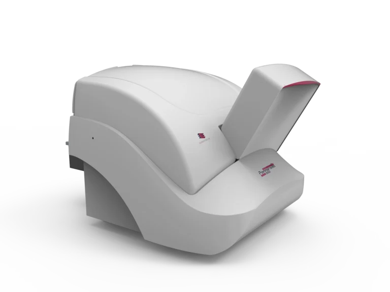
Pannoramic 150 Digital Scanner
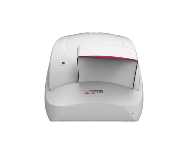
Key Features
- Capacity: Handles 150-slide automatic loading for continuous scanning.
- Automated AI Tissue Detection: Enhances efficiency in biopsy and cytopathology analysis.
- Additional Configuration Option: 3D Peek – Multilayer (Z-Stack) Scanning: Enables in-depth 3D visualization of tumor structures and for optimal cancer diagnostics.
№3. Best for small-to-mid-sized pathology labs with focused oncology workloads.
Pannoramic 75 Digital Scanner

Key Features
- Capacity: Supports up to 75 slides per batch.
- Automated AI Tissue Detection: Enhances efficiency in biopsy and cytopathology analysis.
- Additional Configuration Option: 3D Peek – Multilayer (Z-Stack) Scanning: Enables in-depth 3D visualization of tumor structures and for optimal cancer diagnostics.
№4. Best for medium-to-large hospitals and diagnostic labs managing high biopsy workloads
Pannoramic 250 Flash III DX Digital Scanner
Key Features
- Capacity: Scans up to 300 slides per batch, making it a cost-effective option for large hospitals.
- High-Resolution Brightfield & Cytopathology Scanning: Supports histopathology and cytopathology, crucial for cancer diagnostics.
- AI-Driven Tissue Detection: Fully automated workflow minimizes manual errors.
- Additional customizable option – Multilayer (Z-Stack) & Extended Focus Scanning: Available as an optional configuration, ideal for detailed oncology imaging.
№5. Best for large-scale cancer diagnostics and multi-department pathology institutions
Pannoramic 480 DX Digital Scanner
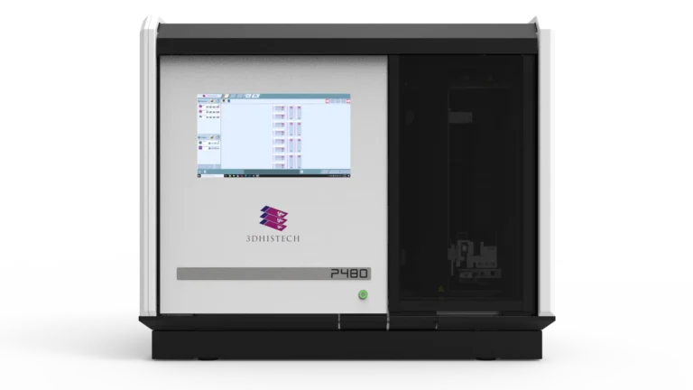
Key Features
- Capacity: Processes up to 480 slides per batch, ensuring uninterrupted scanning.
- Polarization Imaging for Specialized Cancer Diagnostics: Unique feature for detecting birefringent materials (e.g., amyloid plaques, skin cancers).
- Integrated Touchscreen Display: Enables first-level quality control inspections for oncology pathology.
- 10x objective: For large volume scanning in half the time without compromising resolution
- Integrated Z-Stack Imaging for Cytology: Supports multilayer scanning for complex cytopathology cases.
№6. Best for large institutions, research centers, and national pathology networks handling extreme slide volumes
Pannoramic 1000 DX Digital Scanner
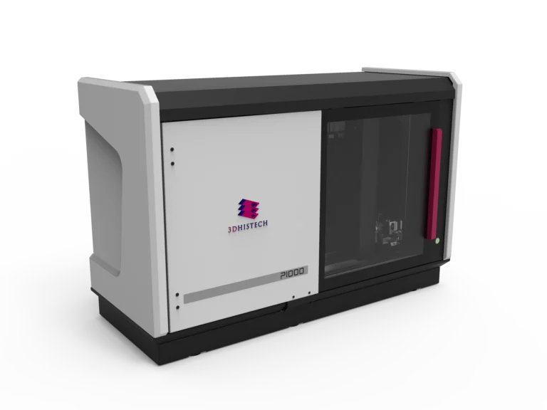
Key Features
- Capacity: Unparalleled 1,200-slide capacity, the highest in the Pannoramic DX series.
- AI-Powered Image Analysis & Automated Workflow: Streamlined batch scanning, minimizing pathologist workload.
- Anti-Vibration Granite Base for Precision Scanning: Ensures high-stability image acquisition for cancer histology and biomarker quantification.
- Seamless Remote Monitoring & Case Integration: Ideal for multi-location pathology institutions requiring networked WSI analysis.
Complete Digital Oncology Pathology Workflow
The Pannoramic DX Series integrates with CaseManager DX, ensuring:
Real-Time Telepathology Consultations
Remote expert collaboration on challenging cancer cases.
Automated Case Management with CaseManager
Assign, track, and review cases within a centralized digital workflow.
AI-Powered Image Analysis with ClinicalViewer and Diagnostic Applications
Quick, Field-of-view-based quantifications and Advanced quantification of tumor markers to assist with treatment decisions.
Do more with the complimentary research software:
Advanced Quantification & AI Analysis with QuantCenter
- AI-Driven Image Analysis: Enables rapid, automated tumor detection.
- Biomarker Quantification: Supports HER2, Ki-67, and PD-L1 scoring in cancer diagnostics.
- High-Speed Server-Based Processing: Handles large datasets efficiently for clinical and research applications
Advanced 3D Microscope Viewer software
3D Reconstruction – For full 3D tissue visualization and analysis. Supports tumor spatial mapping, vascular analysis, and disease progression studies.
Pannoramic DX Digital Scanners for Oncology Pathology Institutions (scanner only)
In large-scale oncology pathology and cancer diagnostics, large laboratories face immense challenges, including high case volumes, the need for ultra-high resolution imaging, and workflow efficiency. The Pannoramic 250 Flash III DX, Pannoramic 480 DX, and Pannoramic 1000 DX Digital Scanners are designed to handle high-throughput scanning in large hospitals, cancer centers, and research institutions. These scanners provide precise, automated, and scalable digital pathology solutions, ensuring fast and accurate whole slide imaging (WSI) for oncology pathology.
By deploying dedicated scanner-only solutions, institutions can integrate these high-volume scanners into their existing LIS, HIS, or telepathology workflows, enabling faster diagnostics, efficient case management, and improved cancer detection.
Why Choose 3DHISTECH’s Pannoramic DX Whole Slide Imaging Solutions?
Ultra-High Resolution for Precise Oncology Diagnostics
Automated, AI-Enhanced Cancer Detection & Quantification
Scalable Solutions for High-Throughput Pathology Labs
Seamless LIS/HIS Integration & Telepathology Capabilities
Regulatory-Compliant Software & Hardware (IVDR-Approved)
Who Benefits from These Telepathology Solutions?
Pathologists & Oncologists
Gain faster, more reliable diagnostic insights with AIassisted scanning and analysis.
Cancer Research Institutions
Enhance quantification and biomarker analysis through high-resolution whole slide imaging.
Hospitals & Diagnostic Labs
Improve turnaround times for biopsy and cytopathology results.
Pharmaceutical & Clinical Trials
Accelerate drug development and precision oncology research.
Telepathology Networks
Facilitate global collaborations and second opinions in cancer cases.