This case study explores how neuroscientists benefit from Pannoramic Confocal, Pannoramic 480, Pannoramic MIDI III, Pannoramic MIDI II, and Pannoramic 150 digital slide scanners, paired with QuantCenter, 3DView, SlideManager, and WebViewer, to address critical challenges in brain tissue analysis, neural imaging, and biomarker discovery.
Key Challenges in Neuroscience Research
Complex 3D Brain Structures
Traditional imaging captures flat, 2D images, limiting the ability to analyze intricate neuronal connections, synaptic density, and cortical layering.
Fluorescence Photobleaching & Sensitivity
Conventional fluorescence imaging degrades signal quality over time, impacting long-term neural protein tracking and multiplex biomarker analysis.
High-Throughput Data Handling
Neuroscience studies generate vast amounts of slide data, requiring efficient scanning, storage, and retrieval to maintain research accuracy and productivity.
Multi-Modal Imaging Needs
Research often involves brightfield, fluorescence, and confocal scanning, necessitating versatile imaging platforms.
Reproducibility & Cross-Laboratory Collaboration
Neuroscientists need standardized imaging and analytical tools to ensure consistent, comparable data across different institutions.
Tissue Microarray (TMA) Integration
Large-scale studies benefit from TMAs, but challenges include variability in sample preparation, data correlation, and spatial analysis of neural tissue biomarkers.
OPTIMAZING Neuroscience RESEARCH
How Whole Slide Imaging Transforms Neuroscience Research
To support geologists, mineralogists, and petroleum researchers, 3DHISTECH offers two cutting-edge imaging solutions:
№1. High-Resolution 3D Imaging of Neural Structures
Pannoramic Confocal Digital Scanner

Why?
Neuroscience demands deep tissue visualization and 3D neural mapping, which the Pannoramic Confocal provides through confocal Z-stack scanning and structured illumination technology.
Real-world Applications
- Neural pathway mapping
Enables neuroscientists to track neuronal connections and
synaptic activity at high resolution. - Neurodegenerative disease studies
Assists in studying diseases like Alzheimer’s and Parkinson’s by imaging amyloid plaques and Lewy bodies with fluorescence multiplexing. - Cellular signaling and neurotransmitter distribution
Provides detailed molecular analysis of dopamine, serotonin, and glutamate receptor distribution in neural tissue.
Software Benefits
- 3DView
Converts Z-stack scans into interactive 3D reconstructions, essential for studying neural circuits. - QuantCenter (PatternQuant, HistoQuant)
Automates pattern recognition and fluorescence quantification, minimizing manual interpretation errors.
№2. High-Throughput Analysis of Large Brain Tissue Sections
Pannoramic 480 Digital Scanner
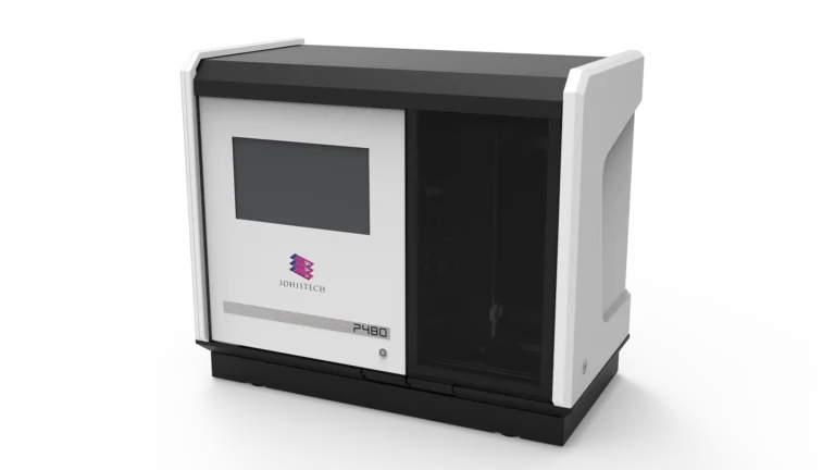
Why?
Neuroscience research requires processing hundreds of brain tissue slides simultaneously, which the Pannoramic 480 excels at with its 480-slide batch capacity and AIpowered scanning.
Real-world Applications
- Comparative brain region studies
Scans multiple slides at once to compare hippocampus, cortex, and basal ganglia in neuroanatomical studies. - Neural stem cell research
Enables the high-throughput imaging of differentiated stem cells for regenerative medicine and neurodevelopmental studies. - Polarization imaging for neurodegeneration
Utilizes polarized light to detect amyloid plaques in Alzheimer’s disease models.
№3. Multi-Modal Imaging for Biomarker Discovery & Disease Research
Pannoramic MIDI III Digital Scanner
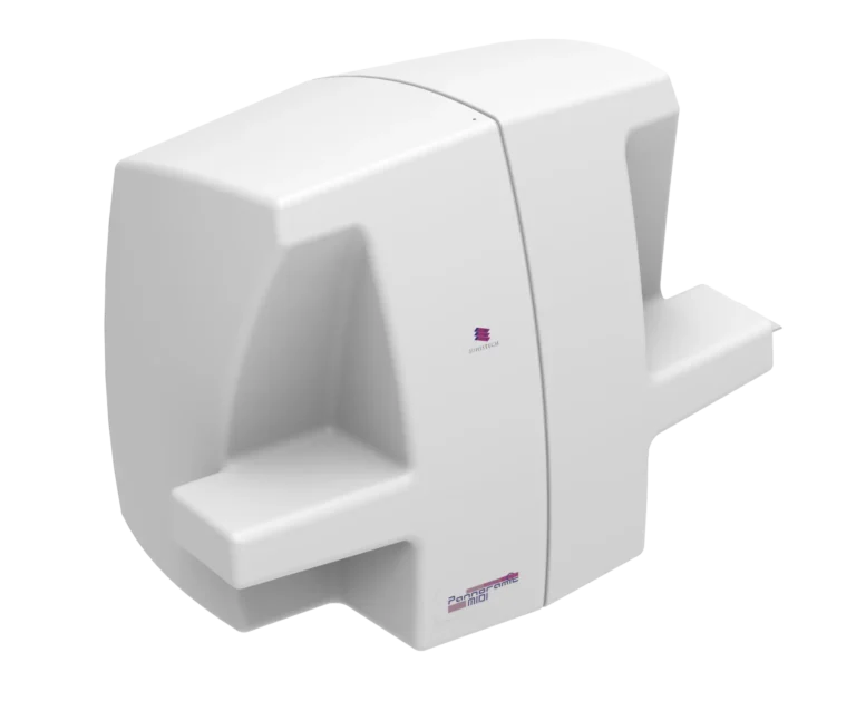
Pannoramic MIDI II Digital Scanner

Why?
Neuroscience research frequently requires multiplex fluorescence imaging, brightfield scanning, and Z-stack analysis.
Real-world Applications
- Brain tumor characterization
Supports multiplex immunofluorescence (IF) and IHC for glioblastoma and metastatic brain cancer analysis. - Neuroinflammation research
Detects inflammatory markers like GFAP and Iba1 in neuroinflammatory conditions. - Toxicology studies
Assesses the impact of pharmaceutical compounds and environmental toxins on neural cells.
Software Benefits
- QuantCenter (HistoQuant, PatternQuant, ScriptQuant)
Provides automated biomarker quantification, improving accuracy and reproducibility. - SlideMaster
Converts scans into various formats for integration with genomics, proteomics, and AI-driven neuroscience analysis platforms.
№4. Fast, High-Resolution Brightfield and Fluorescence Imaging
Pannoramic 150 Digital Scanner
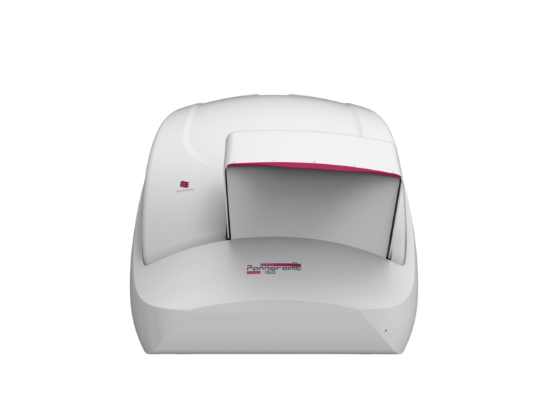
Why?
Neuroscience applications often need fast yet high-resolution brightfield and fluorescence scanning for tissue morphology and cellular analysis.
Real-world Applications
- Rapid brain tissue screening
Quickly analyzes large sections of neural tissue in brain development and aging studies. - Fluorescence imaging for neurotransmitter localization
Scans fluorescence-labeled sections for dopamine and serotonin pathway research. - Neuromuscular disease studies
Helps examine spinal cord degeneration in ALS and muscular dystrophy models.
Software Benefits
- SlideManager
Centralizes neuroscience projects, managing metadata and enabling multi-user collaboration. - WebViewer
Allows remote access for global research teams, improving collaborative multi-center neuroscience studies. - QuantServer
Processes large-scale image analysis in parallel, enhancing speed for high throughput neuroscience research.
OPTIMAZING Neuroscience RESEARCH
Tissue Microarray (TMA) Solutions for Neuroscience Research
To support geologists, mineralogists, and petroleum researchers, 3DHISTECH offers two cutting-edge imaging solutions:
TMA tools:
TMA Master II

TMA Grand Master

TMA molmed

Why?
Neuroscience studies benefit from TMAs by analyzing multiple brain tissue samples on a single slide, reducing variability and increasing efficiency.
Key Challenges Solved
- Standardization
Ensures uniform processing of neural tissue across multiple experimental conditions. - High-throughput biomarker validation
Speeds up studies on neuroinflammation, gliomas, and neurodegeneration. - Reduced costs and sample usage
Maximizes the use of precious brain tissue specimens.
Who Benefits?
- Neuroscientists validating biomarkers in Alzheimer’s, Parkinson’s, and ALS.
- Pharmaceutical researchers testing drug effects on brain tumor samples.
- Neurodevelopmental researchers studying cortical layer formation and synaptic development..
Advancing Neuroscience with Digital Pathology
By integrating high-performance scanners, intelligent software, and TMA solutions, 3DHISTECH’s Whole Slide Imaging platforms revolutionize neuroscience research. Confocal 3D imaging, high-throughput scanning, fluorescence multiplexing, and AI-driven analytics enable neuroscientists to explore brain function, disease mechanisms, and potential therapies with
unprecedented depth and accuracy.
With Pannoramic Confocal, 480, MIDI III, MIDI II, and 150 scanners, supported by QuantCenter, 3DView, SlideManager, and WebViewer, neuroscientists gain the tools needed to transform neurobiology, accelerate discovery, and improve translational research outcomes.