Digital Pathology solutions, powered by 3DHISTECH’s advanced Pannoramic scanners and AI-driven software suite, provide a transformative digital pathology ecosystem for immunology and infectious disease research. These solutions facilitate high-resolution fluorescence imaging, automated biomarker analysis, and 3D tissue reconstruction, enabling researchers to identify immune markers, track infection pathways, and accelerate drug and vaccine development.
Key Challenges in Immunology and Infectious Disease Research
High Complexity in Immunoassays and Biomarker Quantification
Immunohistochemistry (IHC) and multiplex fluorescence imaging require precise marker quantification across multiple tissue regions, but traditional methods lack reproducibility and standardization.
Time-Intensive Image Analysis and Data Interpretation
Manual scoring and annotation of tissue sections introduce subjectivity and variability between researchers.
Limited Sample Throughput for Large-Scale Studies
Epidemiological and vaccine studies require processing and analyzing hundreds to thousands of slides, which is impractical with traditional microscopy.
3D Tissue Architecture and Cellular Interactions Are Difficult to Visualize
Traditional flat, 2D imaging cannot fully capture immune cell interactions, lymphoid structures, or tissue penetration of pathogens.
Data Management and Remote Collaboration Are Challenging
Large image files generated from high-resolution scanning require robust storage, retrieval, and sharing solutions.
3DHISTECH’s Pannoramic digital slide scanners, coupled with AI-powered software and Tissue Microarray (TMA) solutions, overcome these challenges by offering:
High-throughput, ultra-high-resolution imaging with brightfield and fluorescence capabilities
Automated biomarker quantification and AI-assisted analysis for IHC, multiplex fluorescence, and immune profiling.
3D confocal imaging and Z-stack scanning for in-depth tissue structure analysis.
Scalable data management and remote accessibility, supporting global research collaborations.
Optimazing Immunology and Infectious Disease Research
Whole Slide Imaging for Immunopathology and Infectious Disease Research
№1. High-Resolution Whole Slide Imaging Scanners for Immunology & Infectious Disease Research
Pannoramic Confocal Digital Scanner

Best for
Immune cell interaction studies, pathogen tracking, and high-precision fluorescence imaging
Why best?
- Confocal Z-stack scanning enables 3D reconstruction of immune cell architecture in infected tissues
- Minimal photobleaching preserves signal intensity in long-term fluorescence studies.
- Supports multiplex fluorescence for detecting immune markers, cytokines, and viral proteins.
Real-world Application
Studying Immune Response in Viral Infections
Researchers studying SARS-CoV-2 tissue pathology use the Pannoramic Confocal scanner to
visualize infected lung tissues in 3D, identifying T-cell infiltration and inflammatory cytokine
distribution.
Pannoramic 250 FLASH III Digital Scanner

Best for
Large-scale vaccine research, immunohistochemistry (IHC), and biomarker validation.
Why best?
- 45 fluorescence channels allow for high-resolution multiplex biomarker imaging.
- AI-powered automated tissue detection ensures standardized, reproducible analysis.
- 0.12 µm/pixel resolution captures immune cell morphology with subcellular precision.
Real-world Application
IHC Analysis in Tuberculosis Granulomas
Scientists investigating Mycobacterium tuberculosis infection utilize the Pannoramic 250 Flash
III to quantify macrophage and T-cell infiltration across hundreds of granuloma samples in a
high-throughput automated workflow.
Pannoramic MIDI III Digital Scanner
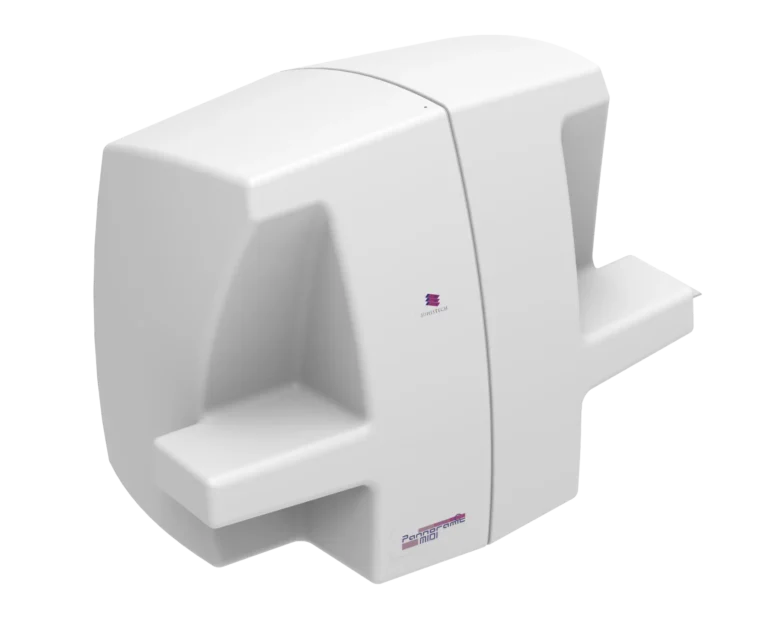
Best for
Biomarker discovery, immune cell profiling, and drug response studies.
Why best?
- Automated switching between brightfield and fluorescence imaging for comprehensive
pathology insights. - AI-assisted biomarker quantification reduces human variability in scoring IHC stains.
- Supports high-content tissue analysis across multiple slides, streamlining drug efficacy studies.
Real-world Application
Tracking Immune Cell Responses in Vaccine Trials
Researchers evaluating mRNA vaccine responses use the MIDI III to analyze CD4/CD8+ T-cell
distribution, helping optimize vaccine formulations based on real-time tissue analysis.
№2. AI-Driven Image Analysis and Workflow Optimization
QuantCenter
AI-Powered Biomarker and Pattern Analysis
- HistoQuant enables precise IHC marker quantification in immune profiling studies.
- PatternQuant identifies immune infiltration patterns in tumor microenvironments and
infectious disease tissues. - AI-based image segmentation enhances the reproducibility of pathogen-induced tissue
damage assessment.
SlideManager
Centralized Image Management for Global Collaboration
- Enables secure, remote access for global teams working on pandemic research.
- Automates data retrieval, annotation, and storage for high-throughput studies.
- Facilitates AI-driven analysis of thousands of whole slide images in centralized repositories.
Real-world Application
Global COVID-19 Pathology Network
Researchers across continents used SlideManager to share and analyze thousands of digitized
COVID-19 lung tissue slides, leading to faster identification of severe inflammatory signatures.
№3. Tissue Microarray (TMA) Solutions for High-Throughput Immunological Research
TMA Master II

TMA molmed

Why?
- TMA compresses hundreds of patient samples into a single slide, reducing reagent and imaging
costs. - Facilitates high-throughput biomarker screening in infectious disease and vaccine research.
- Compatible with AI-powered biomarker quantification in QuantCenter.
Real-world Application
HIV Immunopathology Studies
Scientists analyzing HIV-associated immune dysregulation use TMA molmed to compare immune marker expression across hundreds of patients in a single scanning session, significantly accelerating large-scale studies.
Transforming Immunology and Infectious Disease Research
The Pannoramic WSI ecosystem from 3DHISTECH, including confocal and fluorescence-capable scanners, AI-driven image analysis, and scalable TMA solutions, delivers:
• High-speed, high-precision whole slide imaging for biomarker quantification.
• AI-driven automation to reduce variability and accelerate discoveries.
• 3D imaging capabilities to understand immune cell interactions and pathogen spread.
• Cloud-based collaboration tools for real-time global research.
These advancements revolutionize immunopathology, infectious disease diagnostics, and vaccine development, making breakthroughs faster, more reproducible, and globally scalable.