By integrating fluorescence imaging, confocal scanning, and AI-powered image analysis, these solutions empower laboratories to streamline workflows, ensure reproducibility, and improve diagnostic accuracy. This case study explores how these tools address key challenges in genetics and molecular pathology while transforming research workflows.
Key Challenges in Genetic Research & Digital Imaging
High-Resolution Molecular Imaging Needs
•Researchers require precise fluorescence and brightfield imaging to visualize genetic
markers and molecular interactions.
• Conventional microscopes and manual imaging approaches struggle with consistency
and throughput.
Complexity of Fluorescence & Multiplex Imaging
• Genetic research often involves multiple biomarkers and fluorescence channels,
requiring advanced scanning capabilities.
• Many imaging systems lack the sensitivity and resolution needed for epigenetic and
molecular marker studies.
High-Volume Data Management & Collaboration
• With the increasing scale of genetic studies, efficient data handling and collaboration
tools are crucial.
• Traditional data storage solutions lack the security, accessibility, and scalability needed
for modern genomic research.
Standardization & Reproducibility
• In genetic pathology, accurate quantification of biomarkers and repeatable workflows
are essential for reliable research outcomes.
• Human error and variability in slide scanning pose challenges for consistency.
Advanced Analysis & AI Integration
• Manual interpretation of large-scale molecular images is time-consuming and prone to
bias.
• Researchers need AI-powered quantification tools for automated, standardized
analysis.
Optimizing Genetic Research
Solutions
Each scanner in the Pannoramic series provides unique advantages tailored for molecular cell biology workflows:
№1. 3D Imaging & Z-stack scanning for detailed molecular reconstructions. Ideal for genetic mutation research, epigenetic studies, and biomarker localization. Confocal fluorescence microscopy reduces photobleaching and improves clarity.
Pannoramic Confocal Digital Scanner

№2. Suitable for multiplex biomarker analysis, high-throughput sequencing, and genetic screening. 45 fluorescent channels for high-throughput fluorescence scanning. AI-driven tissue detection & Z-stack scanning for precision.
Pannoramic 250 FLASH III Digital Scanner

№3. Flexible for oncology genetics, pharmacogenomics, and immunogenetics. High-resolution (0.10 µm/pixel) fluorescence & brightfield imaging.12-slide capacity, optimized for biomarker quantification & neuroscience research
Pannoramic MIDI III Digital Scanner
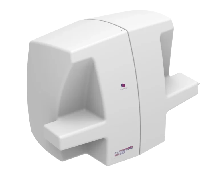
Pannoramic MIDI II Digital Scanner

№4. Efficient for molecular pathology and developmental genetics. Automated barcode reading and Z-stack scanning. Fast fluorescence scanning with a 10x objective for multiplexed imaging.
Pannoramic 150 Digital Scanner
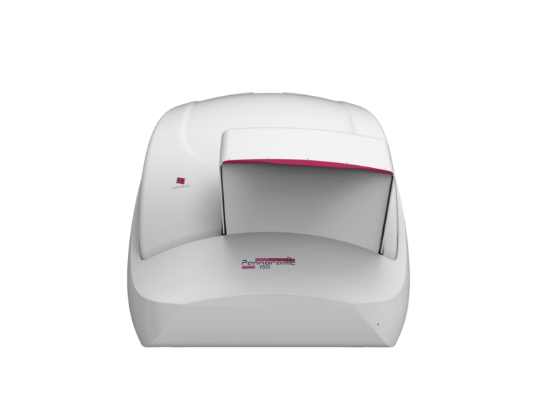
Pannoramic 75 Digital Scanner

№5. Well-suited for toxicogenomics and epigenetic profiling. Fluorescence-specific objectives for in-depth tissue analysis. High-capacity (150 slides), AI-powered fluorescence scanning.
Pannoramic Scan II Digital Scanner

Software Integration for AI-Powered Analysis
To maximize efficiency and accuracy, 3DHISTECH scanners are paired with an advanced software suite:
QuantCenter
AI-driven image quantification for automated biomarker analysis and molecular scoring.
QuantServer
Batch processing and cloud-based computing for handling large datasets in genetics research labs.
SlideManager & SlideCenter
Digital slide management with secure, remote-access capabilities, enabling seamless collaboration in global genomics studies
Applications & Impact
Genetic Mutation Analysis
Using Pannoramic Confocal & QuantCenter, researchers at a molecular genetics institute
analyzed genetic markers in cancerous and non-cancerous tissues.
• Challenge: Distinguishing subtle epigenetic modifications and detecting low-expressing
genetic markers.
• Solution: High-resolution confocal fluorescence imaging provided 3D reconstructions of
mutations, while QuantCenter’s AI analysis quantified expression levels.
• Result: A 30% reduction in analysis time, leading to faster identification of cancer-related
mutations.
Epigenetics & Biomarker Discovery
A pharmaceutical research lab leveraged Pannoramic 250 Flash III & Pannoramic MIDI III for multiplex fluorescence scanning to identify epigenetic biomarkers linked to autoimmune
disorders.
• Challenge: Traditional imaging methods were too slow and lacked the sensitivity needed for
weak fluorescence signals.
• Solution: AI-powered fluorescence scanning (45 channels) delivered sharp, highly detailed
images.
• Result: Enabled more precise biomarker identification, accelerating drug discovery by 40%.
Developmental Genetics & Neuroscience
A university research lab used Pannoramic Scan II for brain tissue analysis in neurodevelopmental disorders.
• Challenge: The team needed high-throughput fluorescent scanning for hundreds of tissue
samples.
• Solution: 150-slide capacity & automated scanning allowed large-scale dataset processing.
• Result: Increased research efficiency by 50%, allowing for faster analysis of neurogenetic
pathways.
Tissue Microarrays (TMA) in Genetic Research
TMA molmed & TMA Master II – Precision & Efficiency
• TMA molmed: High-throughput tissue sampling, ideal for PCR, dPCR, and sequencing
applications oai_citation:8‡Tissue Microarray Solutions with the TMA molmed.docx.
• TMA Master II: Compact, fully automated TMA creation, enabling up to 250 cores per hour,
ensuring high sample reproducibility oai_citation:9‡Tissue Microarray Solutions with the TMA Master II.docx.
Why TMAs Matter in Genetics
• Challenge: Large-scale genetic studies require consistent, reproducible tissue samples for
biomarker validation.
• Solution: Automated TMA creation & digital integration reduce sample variability and
improve standardization.
Who Benefits?
• Biopharmaceutical companies developing targeted gene therapies.
• Genomics research centers conducting large-scale biomarker screenings.
• Hospitals & clinical trials validating new genetic diagnostic tools.
3DHISTECH’s Pannoramic Digital Pathology solutions, coupled with QuantCenter, SlideManager, and advanced tissue microarray tools, revolutionize genetic research workflows. By offering high-resolution imaging, AI-powered analysis, and efficient tissue microarraying, these solutions enhance accuracy, reduce processing time, and improve biomarker quantification.
Key Benefits:
- Ultra-high-resolution imaging for precise genetic marker visualization.
- Advanced fluorescence scanning (up to 45 channels) for multiplexed molecular studies.
- AI-driven quantification eliminates manual errors and improves standardization.
- Tissue microarray integration ensures high-throughput, reproducible studies.
- Cloud-based data management & collaboration tools streamline large-scale genomics research.
By integrating these technologies, research institutions and pharmaceutical companies can accelerate genetic discoveries, drive precision medicine, and enhance translational research outcomes.