This case study explores how 3DHISTECH’s Whole Slide Imaging (WSI) and 3D Digital Pathology solutions—including the Pannoramic Confocal, Pannoramic MIDI III, Pannoramic Scan II, Pannoramic 150, and Pannoramic-X Micro-CT Scanner—provide an advanced platform to overcome these challenges. It also highlights how AI-powered QuantCenter, SlideViewer, WebViewer, and CT Control & 3DView software facilitate deep biological insights.
Key Challenges in Developmental Biology Research
Limited Imaging Resolution & Depth
Traditional microscopy methods capture only 2D slices of tissues, making it difficult to study 3D cellular structures and interactions.
Time-Consuming & Inconsistent Image Analysis
Manual slide scanning and analysis are slow and subject to variability, making it difficult to track longitudinal changes in cell differentiation.
Difficulty in Biomarker Quantification
Analyzing molecular markers across multiple layers and fluorescence channels is challenging without AI-driven image processing.
Inability to Study Whole Tissues Without Sectioning
Conventional histology requires physical sectioning, which can distort tissue morphology and cause sample loss.
Collaboration and Data Management Issues
Large-scale research projects require efficient digital storage, real-time data sharing, and multi-user access, which traditional methods struggle to support.
Optimizing Developmental Biology
Solution Implementation: Integrating 3DHISTECH’s Digital Pathology Technology
№1. High-Resolution Whole Slide Imaging with Pannoramic Scanners
Researchers at a developmental biology lab studying stem cell differentiation integrated Pannoramic Confocal, MIDI III, Scan II, and 150 Digital Scanners into their workflow.
What Scanners provided:
• Confocal Z-stack scanning for multi-layered imaging and 3D reconstructions (Pannoramic Confocal).
• High-resolution fluorescence and brightfield imaging (Pannoramic MIDI III, Scan II, and 150).
• Rapid automated scanning of up to 150 slides for large-scale developmental studies (Pannoramic Scan II & 150).
Key Benefits
- Enhanced 3D visualization of developing tissues.
- Faster, automated high-throughput scanning reduced workflow time.
- Fluorescence imaging allowed biomarker tracking across multiple channels.
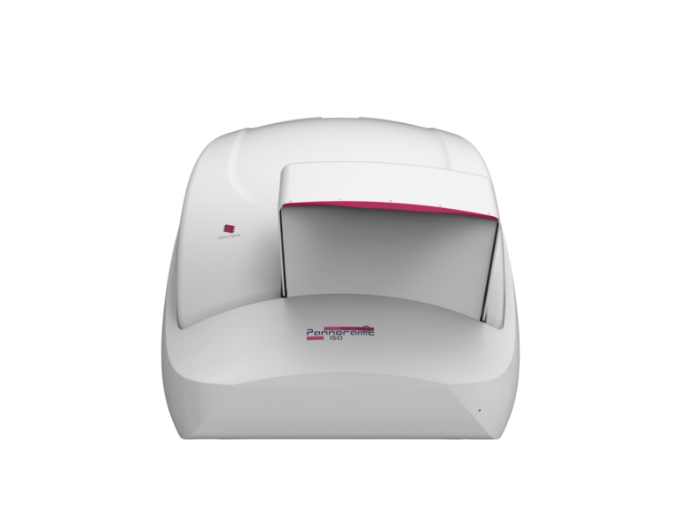
Pannoramic 150 Digital Scanner
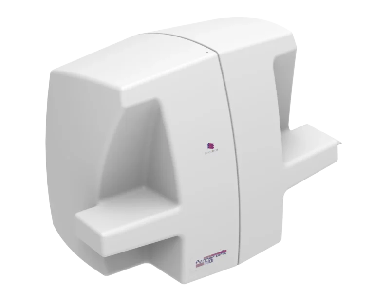
Pannoramic MIDI III Digital Scanner
Pannoramic Scan II Digital Scanner

Pannoramic Confocal Digital Scanner

№2. AI-Powered Image Analysis & Biomarker Quantification
The lab deployed QuantCenter software, which includes PatternQuant (for pattern recognition), HistoQuant (for quantification of tissue structures), and NuclearQuant (for cell-based analysis).
Real Scenario
A research group analyzing gene expression in neural crest cells used HistoQuant to quantify fluorescence signals from multiple markers. This automated method replaced manual counting, reducing analysis time from weeks to hours.
Key Benefits
- AI-powered automated quantification eliminated manual subjectivity.
- Faster, more reproducible results for biomarker validation.
- Multi-channel fluorescence analysis improved tracking of developmental stages.
№3. 3D Digital Pathology with Pannoramic-X Micro-CT
To study tissue organization without sectioning, researchers used the Pannoramic-X Micro-CT scanner.
This allowed for:
- Virtual slicing and staining of whole tissue blocks.
- High-resolution 3D reconstructions of embryonic and stem cell structures.
- Quantitative biomarker analysis for tumor development and tissue regeneration studies.
Real Scenario
A team studying heart tissue regeneration after injury used the Pannoramic-X to analyze blood vessel formation in 3D, without having to destroy the sample.
Key Benefits
- Non-destructive imaging preserved tissue integrity.
- 3D visualization allowed for deeper insights into morphological changes.
- Virtual staining eliminated the need for physical sectioning and reprocessing.
Pannoramic-X

№4. Enhancing Collaboration & Data Management
To streamline data sharing, SlideManager, WebViewer, and SlideViewer were implemented, enabling:
- Remote access for global research teams.
- Secure storage of large datasets.
- Multi-slide comparisons for tracking developmental changes over time.
Real Scenario
A multi-institutional study on fetal tissue development used WebViewer to allow researchers across different locations to collaborate in real-time, comparing images and annotating tissue structures simultaneously.
Key Benefits
- Seamless remote collaboration across research centers.
- Efficient data storage & retrieval improved workflow speed.
- Multi-slide comparisons enhanced cross-sectional studies of tissue samples.
Results & Impact
Higher Throughput & Faster Research Progress
Automated scanning and AI-driven analysis reduced experiment times by 50%.
Deeper Insights with 3D Imaging
Confocal and Micro-CT imaging enabled detailed structural studies, enhancing tissue visualization.
Improved Biomarker Validation & Quantification
AI-driven analysis provided more accurate and reproducible biomarker data.
Better Collaboration & Accessibility
Cloud-based solutions allowed instant access to scanned slides, improving international research efforts.
Who Benefits from These Solutions?
Developmental Biologists
Gain access to high-resolution imaging, fluorescence & 3D tissue analysis to study cell differentiation and organ development.
Stem Cell Researchers
Can track stem cell differentiation in 3D, leading to insights into regenerative medicine.
Geneticists
Use AI-powered biomarker quantification to correlate genetic mutations with developmental changes.
Neuroscientists
Study brain tissue development with multi-layer fluorescence scanning and micro-CT imaging.
Pharmaceutical Companies
Use AI-powered analytics for drug development, especially in regenerative therapies.
The integration of 3DHISTECH’s Whole Slide Imaging and 3D Digital Pathology solutions transforms Developmental Biology research by providing high-resolution, automated, and AI-powered tools. These innovations solve the key challenges of limited imaging resolution, slow analysis, biomarker quantification, and collaboration inefficiencies.
By implementing these solutions, researchers accelerate discoveries, improve accuracy, and enhance scientific collaboration, driving forward advances in stem cell research, organ development, and regenerative medicine.