This case study highlights how 3DHISTECH’s Pannoramic Digital Pathology solutions, Tissue Microarray (TMA) technologies, and Pannoramic-X Micro-CT scanning address key challenges in cancer, immunology, and infectious disease research, providing researchers with an integrated digital pathology workflow for precision oncology and translational medicine.
Key Challenges in Cancer Research & Digital Pathology
Need for High-Throughput, High-Resolution Imaging
• Challenge: Cancer studies require high-throughput scanning of large tissue samples while maintaining superior resolution and fluorescence multiplexing.
• Solution: The Pannoramic Confocal, Scan II, 480, MIDI III, and Pannoramic 150 Digital Scanners provide high-resolution, multi-channel fluorescence, and confocal Z-stack imaging, enabling detailed analysis of tumor microenvironments, genetic mutations, and immune cell infiltration.
Limitations of Traditional Tissue Analysis
• Challenge: Conventional histology relies on 2D slides, which limit spatial understanding of tumor morphology and biomarker localization.
• Solution: The Pannoramic-X Micro-CT Scanner introduces 3D digital pathology, allowing virtual slicing and volumetric tumor assessment without physical sectioning.
Inefficiencies in Biomarker Quantification
• Challenge: Manual immunohistochemistry (IHC) interpretation is time-consuming, subjective, and prone to inter-observer variability.
• Solution: QuantCenter AI-powered analysis enables precise biomarker quantification, pattern recognition, and automated IHC scoring.
Data Management & Global Collaboration
• Challenge: Research institutions struggle with large-scale digital slide management, remote access, and data sharing.
• Solution: SlideManager & SlideCenter provide centralized data management and cloud-based sharing, fostering collaboration across research centers.
Advancing Immunology & Infectious Disease Research
• Challenge: Studying immune responses to tumors and infectious diseases requires multiplex imaging and high-throughput tissue sampling.
• Solution: TMA molmed & TMA Master II tissue microarray solutions accelerate large-scale biomarker studies, vaccine development, and personalized medicine research.
№1. High-Throughput, High-Resolution Imaging
Best for 3D fluorescence imaging of tumor samples:
Pannoramic Confocal Digital Scanner

Real-World Application
A research team studying tumor microenvironments uses the Pannoramic Confocal scanner to create 3D reconstructions of tissue sections, identifying hypoxic tumor regions and immune cell interactions critical for cancer immunotherapy
Optimized for immunohistochemistry (IHC) and fluorescence research:
Pannoramic Scan II Digital Scanner

Ideal for high-throughput cancer research (1200 & 480-slide capacity):
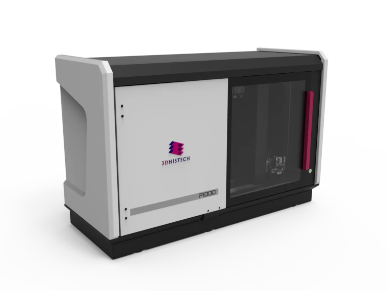
Pannoramic 1000 Digital Scanner
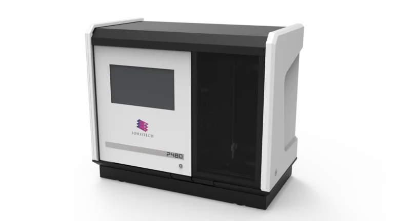
Pannoramic 480 Digital Scanner
Versatile solutions for biomarker discovery & multiplex imaging:
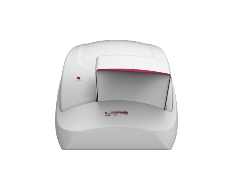
Pannoramic 150 Digital Scanner
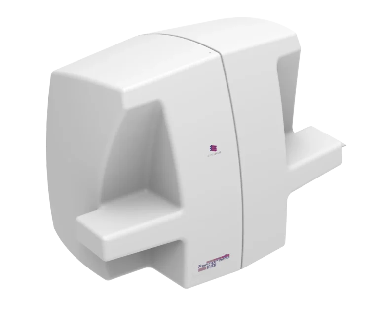
Pannoramic MIDI III Digital Scanner
№2. AI-Driven Biomarker Quantification & Tumor Profiling
Software Solutions
Real-World Application
In a lung cancer clinical trial, researchers use QuantCenter’s HistoQuant module to analyze PD-L1 expression in IHC-stained sections, ensuring accurate patient stratification for immunotherapy.
№3. Tissue Microarray (TMA) for Large-Scale Biomarker Studies
Key Challenges in Large-Scale Cancer & Infectious Disease Research
- Manual biomarker validation is time-consuming & resource-intensive.
- High-throughput screening of tissue samples is essential for vaccine & drug discovery.
What we offer
- Automated microarraying of FFPE tissue samples, reducing hands-on time.
- Supports multiplex biomarker analysis, enabling high-density tissue sampling.
- Ideal for epidemiological studies, vaccine development, and precision medicine research.
TMA Master II

TMA Grand Master

TMA molmed

Real-World Application
A research team studying COVID-19 vaccine responses uses TMA molmed to extract tissue microarrays from infected lung biopsies, comparing cytokine expression across multiple patients.
№4. Pannoramic-X Micro-CT: A New Frontier in 3D Digital Pathology
Why 3D Imaging Matters in Cancer Research
- Traditional histology provides only 2D snapshots of tumor architecture.
- Understanding tumor morphology, lymphovascular invasion, and biomarker distribution requires 3D visualization.
Transforming Tumor Analysis
- Non-destructive, whole-block 3D imaging of tumor samples.
- Virtual slicing & staining, eliminating the need for sectioning.
- AI-powered volumetric analysis for biomarker quantification & treatment response evaluation.
Real-World Application
A breast cancer research institute uses Pannoramic-X to analyze HER2 expression in full tumor blocks, uncovering previously undetected microinvasions, improving treatment decisions.
How This Solution Advances Cancer & Immunology Research
Higher Throughput
Scanners like Pannoramic 480 & Scan II enable massive sample digitization.
Deeper Insights
Pannoramic Confocal & Pannoramic-X offer 3D tissue visualization & quantitative analysis.
AI-Powered Quantification
QuantCenter & TMA molmed ensure precise biomarker measurements.
Scalable & Collaborative
SlideManager & SlideCenter allow global research collaboration & data integration.
By combining WSI, AI-driven analysis, TMA technology, and micro-CT scanning, 3DHISTECH’s digital pathology ecosystem revolutionizes cancer research, accelerating breakthrough discoveries in oncology, immunology, and infectious diseases.
Who Benefits from These Solutions?
Oncology Research Centers
High-throughput biomarker validation & tumor profiling.
Pharmaceutical Companies
Faster drug discovery & clinical trials.
Immunologists & Infectious Disease Specialists
Multiplex biomarker analysis for immune response & vaccine research.
Pathologists & Translational Researchers
AI-driven quantitative pathology with WSI & microCT integration.