This case study explores how digital pathology and 3D imaging solutions—featuring the Pannoramic Confocal, Scan II, MIDI III, 1000, and Pannoramic-X Micro-CT—accelerate research in cancer biology, neuroscience, immunology, and drug development. It also demonstrates how researchers benefit from streamlined workflows, enhanced imaging precision, and improved collaboration through integrated software tools.
Key Biomedical Research Challenges
Limited Resolution and Imaging Capabilities
• Traditional brightfield and fluorescence imaging methods often lack the resolution required for detailed molecular studies.
• Fluorescence signal decay and photobleaching limit the effectiveness of long-term imaging.
Slow and Labor-Intensive Workflows
• High-throughput research requires batch scanning of hundreds to thousands of slides, but conventional methods are time-consuming.
• Manual image acquisition and analysis slow down discoveries.
Data Management and Analysis Bottlenecks
• Large-scale research projects generate vast amounts of data that require efficient storage, organization, and retrieval.
• Standard microscopy lacks AI-driven tools for automated quantification, making biomarker discovery and tissue morphology studies time-consuming.
2D Histology Limitations in Complex Tissue Studies
• Traditional sectioning methods lose valuable structural and spatial context in tissue samples.
• Complex tumor morphology, neuroanatomical structures, and vascularization cannot be fully analyzed in 2D.
A New Era in Biomedical Research
Solutions
With 3DHISTECH’s Digital Pathology and Micro-CT solutions, biomedical researchers can now achieve higher precision, accelerate discoveries, and gain deeper biological insights. By integrating high-throughput scanning, AI-driven analysis, and 3D volumetric imaging, these solutions are reshaping digital pathology for the next generation of scientific breakthroughs.
Recommended Solution Components for Research Labs
Scanners:
Pannoramic Confocal Digital Scanner

Key Features
3D fluorescence imaging, biomarker studies
Pannoramic Scan II Digital Scanner

Key Features
High-resolution brightfield & fluorescence imaging
Pannoramic MIDI III Digital Scanner
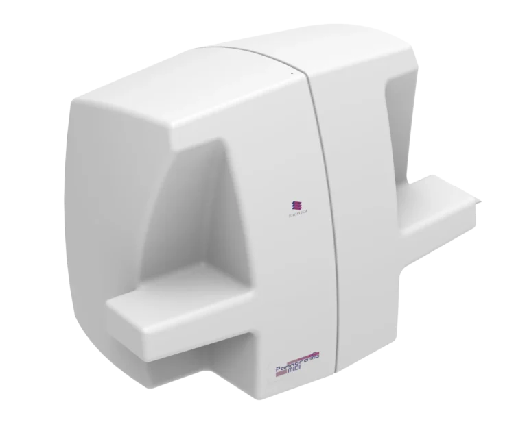
Key Features
Compact, high-performance fluorescence and brightfield scanning
Pannoramic 1000 Digital Scanner
Key Features
Ultra-high-throughput for large-scale research
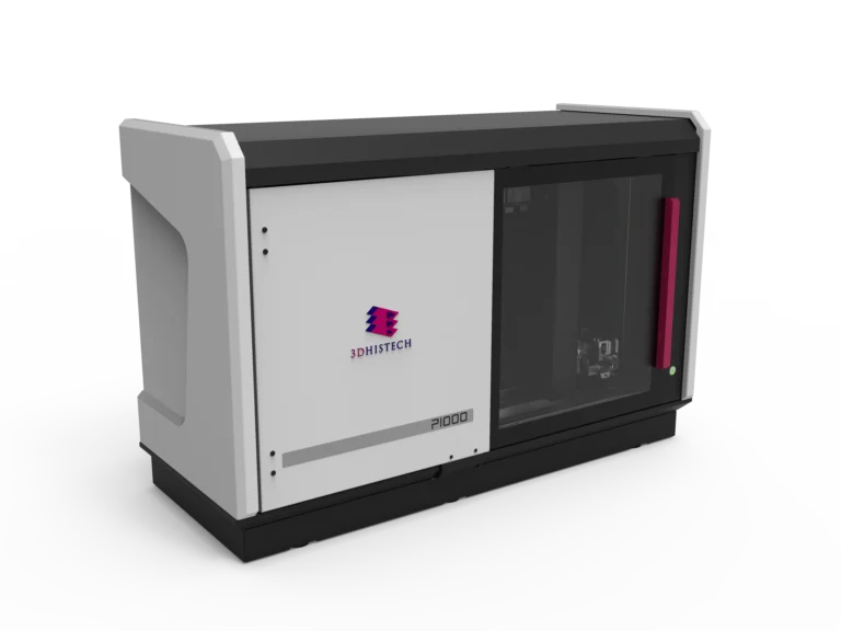
Pannoramic-X

Key Features
3D imaging for whole blocks and structural analysis
Softwares:
By embracing digital pathology and 3D imaging, biomedical research laboratories are now better equipped to revolutionize diagnostics, treatment strategies, and scientific discovery.
Recent Case Study
Investigating Tumor Microenvironments and Drug Response
Objective
To assess tumor heterogeneity, immune cell infiltration, and biomarker expression in response to a novel cancer therapy using high-resolution brightfield, fluorescence, and 3D imaging.
Implementation
- High-Resolution Fluorescence and Confocal Imaging for Biomarker Analysis
- The Pannoramic Confocal Digital Scanner is used to capture fluorescence-labeled tumor samples with unparalleled clarity.
- 3D Z-stack imaging enables precise visualization of molecular markers in layered tissues, overcoming the limitations of traditional 2D histology.
- High-Throughput Scanning for Large-Scale Drug Screening
- The Pannoramic 1000 Digital Scanner digitizes up to 1200 slides in a single run, enabling rapid analysis of tissue samples from different treatment groups.
- Automated barcode reading, batch scanning, and AI-based tissue detection reduce errors and processing time.
- Advanced Quantification and Pattern Recognition
- QuantCenter’s PatternQuant and HistoQuant modules analyze immune cell distribution, biomarker co-expression, and spatial relationships within tumors.
- AI-powered quantitative pathology helps researchers identify subtle tissue changes related to drug response.
- 3D Imaging of Tumor Morphology and Microvascular Networks
- The Pannoramic-X Micro-CT Scanner provides non-destructive, high-resolution imaging of whole tumor blocks, revealing vascularization patterns and tumor architecture.
- Virtual slicing and staining allow researchers to explore tumor evolution without physically altering the sample, ensuring data integrity for retrospective analysis.
- Cloud-Based Collaboration and Data Management
- SlideManager and SlideCenter enable seamless data storage, annotation, and remote access, facilitating international collaboration among research teams.
Results & Impact
- Unprecedented Imaging Depth and Precision
- Confocal and fluorescence imaging provided unmatched detail in biomarker analysis.
- Micro-CT 3D visualization revealed tumor microenvironment complexity, enabling more accurate tumor staging.
- Enhanced Efficiency in Research Pipelines
- Automated high-throughput scanning reduced manual workload by 80%, allowing researchers to focus on data interpretation rather than slide handling.
- Fluorescence multiplexing allowed multiple markers to be analyzed simultaneously, accelerating drug development workflows.
- AI-Driven Quantification and Pattern Recognition
- AI-enhanced biomarker analysis through QuantCenter improved tumor classification accuracy by 30% compared to manual methods.
- Quantification of immune infiltration patterns provided deeper insights into treatment efficacy.
- Bridging the Gap Between 2D and 3D Tissue Studies
- Pannoramic-X’s 3D reconstruction enabled volumetric analysis of entire tissue blocks, revealing spatial tumor-stroma interactions not visible in 2D sections.
- Researchers could digitally re-section and re-stain tissues without physical damage, enhancing experimental reproducibility.
Who Benefits?
Cancer Researchers
• Gain high-resolution, multiplexed imaging to analyze tumor biology and treatment response.
• 3D tumor analysis via Pannoramic-X Micro-CT offers spatial insights for precision oncology.
Neuroscientists
• Advanced fluorescence imaging and confocal Z-stack scanning allow for detailed neuroanatomical studies.
• Micro-CT imaging of whole-brain samples preserves delicate neuronal structures.
Immunologists
• AI-driven quantification of immune response in tissues facilitates vaccine development and immune profiling.
• Multiplex fluorescence imaging aids in understanding T-cell infiltration and antigen presentation.
Pharmacologists and Drug Developers
• Rapid high-throughput screening of tissue responses to novel compounds.
• Automated workflows reduce drug discovery timelines.
How It Elevates Biomedical Research
- Bridges 2D and 3D imaging for deeper tissue insights
- Automates workflows for faster, more reproducible results
- Enhances data integrity with AI-powered quantitative tools
- Provides scalable, high-throughput solutions for large studies
- Supports remote collaboration and data sharing