This case study explores how 3DHISTECH’s digital imaging and AI-driven analysis tools enhance fossil and artifact research by preserving structural integrity while enabling deeper, high-throughput scientific inquiry.
Key Challenges in Archaeology and Paleontology Research
Non-Destructive Analysis of Fossils & Artifacts
• Traditional histology and sectioning risk damaging rare or fragile specimens.
• Some artifacts and fossils are too small for macroscopic analysis yet too fragile for standard thin-sectioning techniques.
Revealing Microstructures in Fossilized Remains & Minerals
• Ancient tissues and mineral compositions are not always visible using standard brightfield microscopy.
• Fossilized bone remodeling, growth rings, and diagenetic changes require multi-layer scanning and high-magnification imaging.
Analyzing & Preserving Large Specimens
• Many fossils exceed the size limits of conventional slide scanners, making 3D digital imaging essential.
• Paleontological specimens often contain complex, multi-layered structures that require virtual sectioning without physical alteration.
Identifying Organic Residues & Ancient Pigments
• Soft tissue residues, pigments, and biomineralization processes require specialized imaging modalities such as polarization and fluorescence microscopy.
• Detecting ancient organic matter in fossils can provide new insights into evolutionary biology.
Enhancing Global Scientific Collaboration
• Research on rare artifacts and fossils is often limited to institutions that physically possess the specimens.
• High-resolution digital imaging with remote access capabilities enables cross-institutional comparative studies without shipping or handling delicate specimens.
Optimizing Archaeology & Paleontology research
Solutions
Scientific Solutions for Digital Paleontology & Archaeology Research
№1. Pannoramic-X Micro-CT Scanner
Key Benefits
• Non-destructive 3D digital imaging of fossils, bones, and artifacts using soft X-ray micro-CT technology.
• Virtual slicing and staining, enabling detailed internal structure analysis without physical alterations.
• Supports large specimens up to 15 cm, allowing detailed visualization of skeletal microstructures, diagenetic changes, and fossil inclusions.
• AI-driven pattern recognition for automated analysis of tissue composition and mineralization patterns.
Pannoramic-X

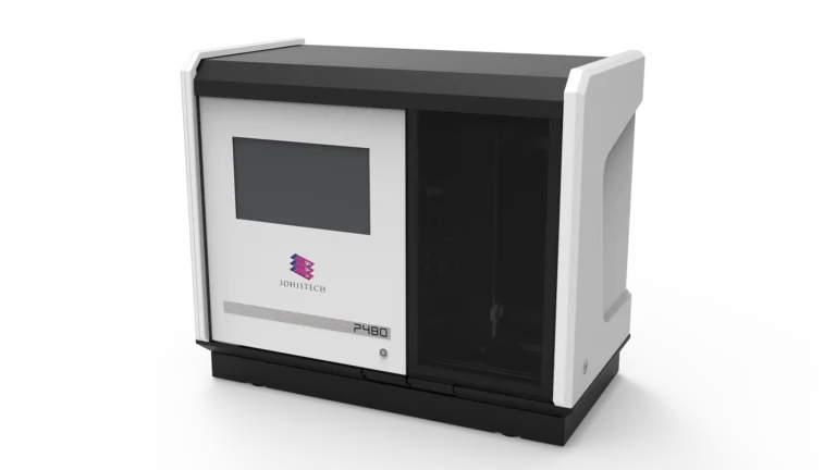
№2. Pannoramic 480 Digital Scanner (with Polarization Imaging & High-Throughput Capabilities)
Key Benefits
• Polarized light microscopy to analyze birefringent structures such as mineralized fossils, crystalline formations, and ancient pigments.
• Z-stack multi-layer imaging, providing depth-enhanced scans of fossilized bone histology and sedimentary microstructures.
• High-throughput scanning of up to 480 slides, enabling large-scale analysis of thin-sectioned fossils and artifacts.
Pannoramic 480 Digital Scanner
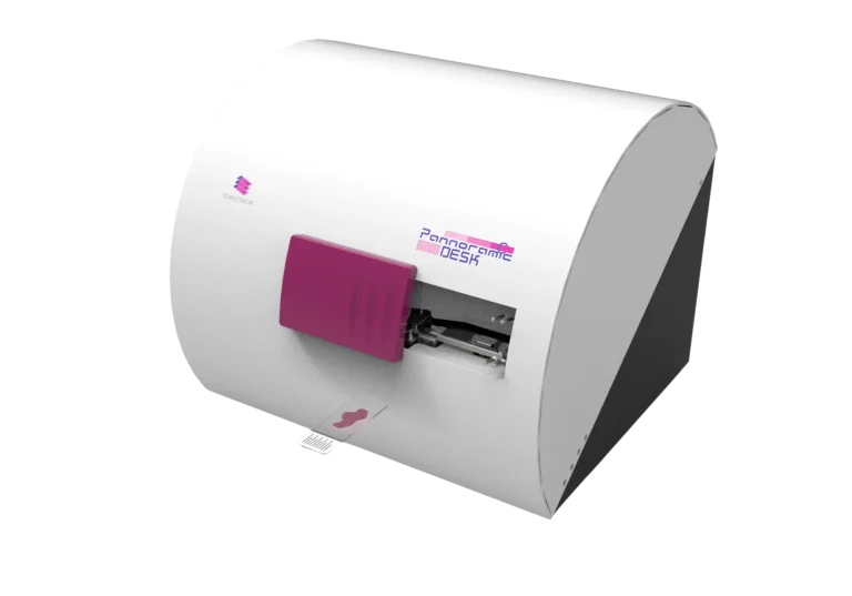
№3. Pannoramic Desk II DW Digital Scanner
Key Benefits
• Brightfield whole-slide scanning for high-resolution thin-sectioned archaeological samples.
• Double-wide slide support, accommodating large-format thin sections commonly used in paleontological petrography.
• Optional Z-stack scanning for multilayer analysis, improving visualization of microfossils and cellular remnants.
Pannoramic Desk II DW Digital Scanner
№4. QuantCenter AI Image Analysis
Key Benefits
• PatternQuant AI detects microstructural patterns in fossils, including growth rings, collagen degradation, and mineral deposits.
• Automated segmentation of fossilized tissues and mineral inclusions, reducing manual annotation variability.
• Quantitative biomarker analysis, aiding in the comparative classification of fossils across different geological epochs.
№5. SlideViewer & WebViewer
Key Benefits
- Remote access to digitized fossil and artifact slides, allowing international researchers to collaborate without handling the original specimens.
- Multi-slide comparison and annotation tools, facilitating cross-institutional research on ancient biological specimens.
SlideViewer & WebViewer
Remote collaboration & multi-slide visualization
Recent Case Study
High-Resolution Digital Analysis of a Neanderthal Fossil
Scenario
A multinational research team seeks to analyze a 50,000-year-old Neanderthal femur to determine:
- Bone density and mineralization patterns over time.
- Possible collagen remnants and soft tissue structures preserved in fossilized bone.
- Evidence of stress fractures and bone remodeling, indicating lifestyle and environmental adaptations.
Implementation
- Pannoramic-X Micro-CT Scanner is used to generate 3D reconstructions of the femur, providing virtual cross-sections to study internal bone density variations and cortical remodeling.
- Pannoramic 480 Digital Scanner (Polarization Imaging) detects birefringent structures, potentially indicating collagen residues or ancient mineral deposits.
- QuantCenter AI automatically analyzes bone microstructure patterns, identifying diagenetic changes and remodeling indicators.
- SlideViewer enables international research teams to collaborate remotely, with real-time annotations and comparative analysis.
Results & Impact
- High-resolution, non-destructive fossil imaging, preserving the specimen for future studies.
- Detection of subtle bone remodeling markers, suggesting Neanderthal lifestyle adaptations.
- Identification of potential collagen remnants, opening new avenues for ancient biomolecular studies.
- Standardized, AI-powered analysis, reducing human error in fossil classification.
- International research collaboration without physical specimen handling, accelerating peer-reviewed studies.
Who Benefits and How?
Paleoanthropologists & Evolutionary Biologists
• Reconstruct hominin lifestyles by analyzing bone microstructure, collagen preservation, and stress fracture patterns.
• Compare fossilized remains across different populations, revealing migration and adaptation patterns.
Archaeological Research Institutions
• Enable early and accurate detection of malignancies through ultra-resolution imaging.
• Provide quantitative analysis to support diagnostic confidence.
Museum Research Laboratories
• Preserve and study historical artifacts without degradation, maintaining collections for future generations.
• Create digital archives of ancient specimens, facilitating virtual exhibits and research collaborations.
Geological & Environmental Scientists
• Analyze mineralization processes in fossils, contributing to knowledge of paleoenvironmental conditions.
• Track diagenetic changes in archaeological materials, helping reconstruct historical climates and ecosystems.
A New Era in Archaeology and Paleontology
The integration of whole slide imaging, micro-CT scanning, AI-driven analysis, and digital collaboration tools is transforming paleontological and archaeological research.
- Pannoramic-X and Pannoramic 480 provide non-destructive, high-resolution imaging of fossils and artifacts, preserving them for future study.
- Polarization and Z-stack imaging reveal previously undetectable microstructures, improving artifact and fossil classification.
- QuantCenter AI accelerates fossil analysis, ensuring reproducible and objective results.
- Digital slide-sharing tools enable global scientific collaboration, reducing the need for physical specimen transport.
By adopting these state-of-the-art digital pathology solutions, the scientific community can conduct more accurate, scalable, and sustainable research, ensuring the preservation of our biological and cultural heritage for future generations.