QuantCenter offers modular, high-precision image analysis for whole-slide
quantification in histopathology and molecular pathology. Unlike most
quantification software, it is more flexible than one-size-fits-all solutions with
customizable modules tailored to specific analysis needs, features customizable AIpowered segmentation and deep-learning capabilities delivering superior
accuracy, multi-slide batch processing, and integrated visualization tools which
provide comprehensive insights without requiring external software. All these
offers maximum flexibility for researchers and its scalable architecture make it the
ultimate solution for quantitative pathology research.
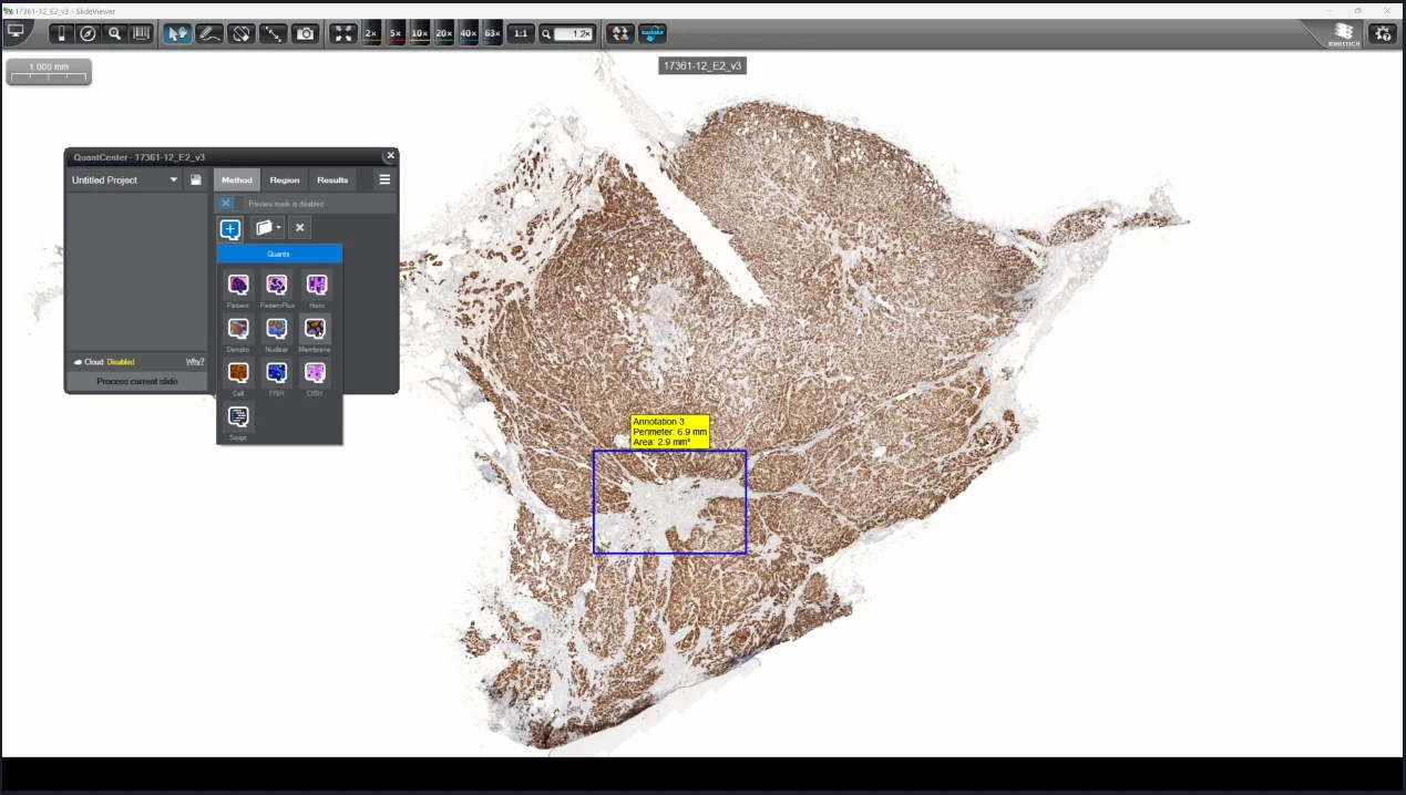
QuantCenter is an image analysis platform for whole-slide quantification in histopathology and molecular pathology. With customizable algorithms and linkable modules. Integrated with SlideManager, it enables efficient execution of analysis on research study samples, driving precise reproducible results. It offers a range of combinable modules including:
HistoQuant
Identifies stained tissue elements based on color and intensity features.
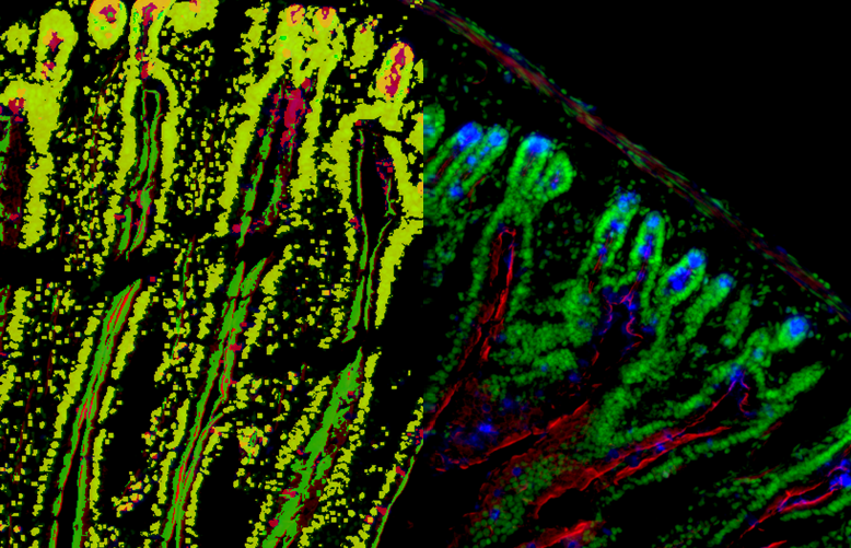
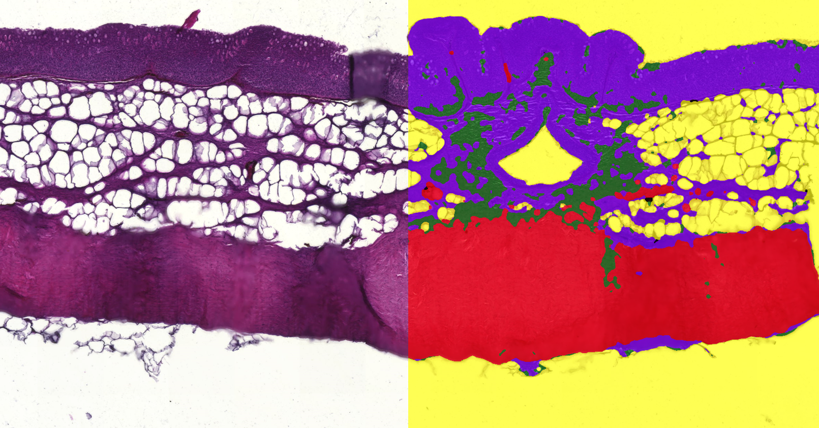
Elongated villous structure with multi-layered nuclei analysis
by PatternQuant
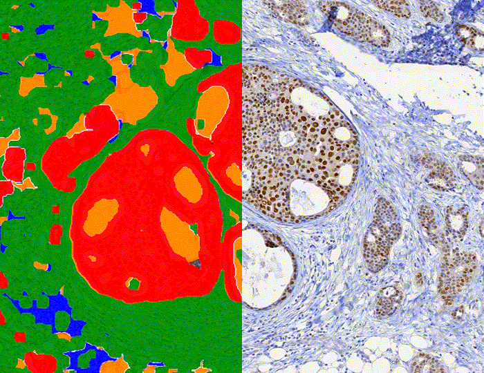
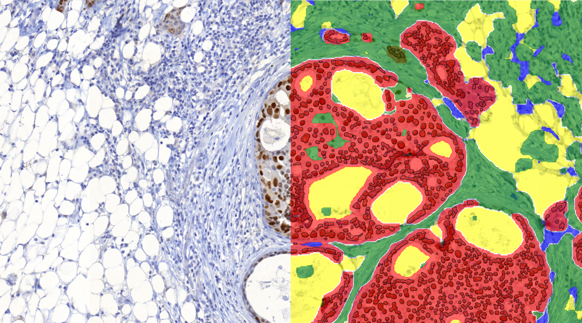
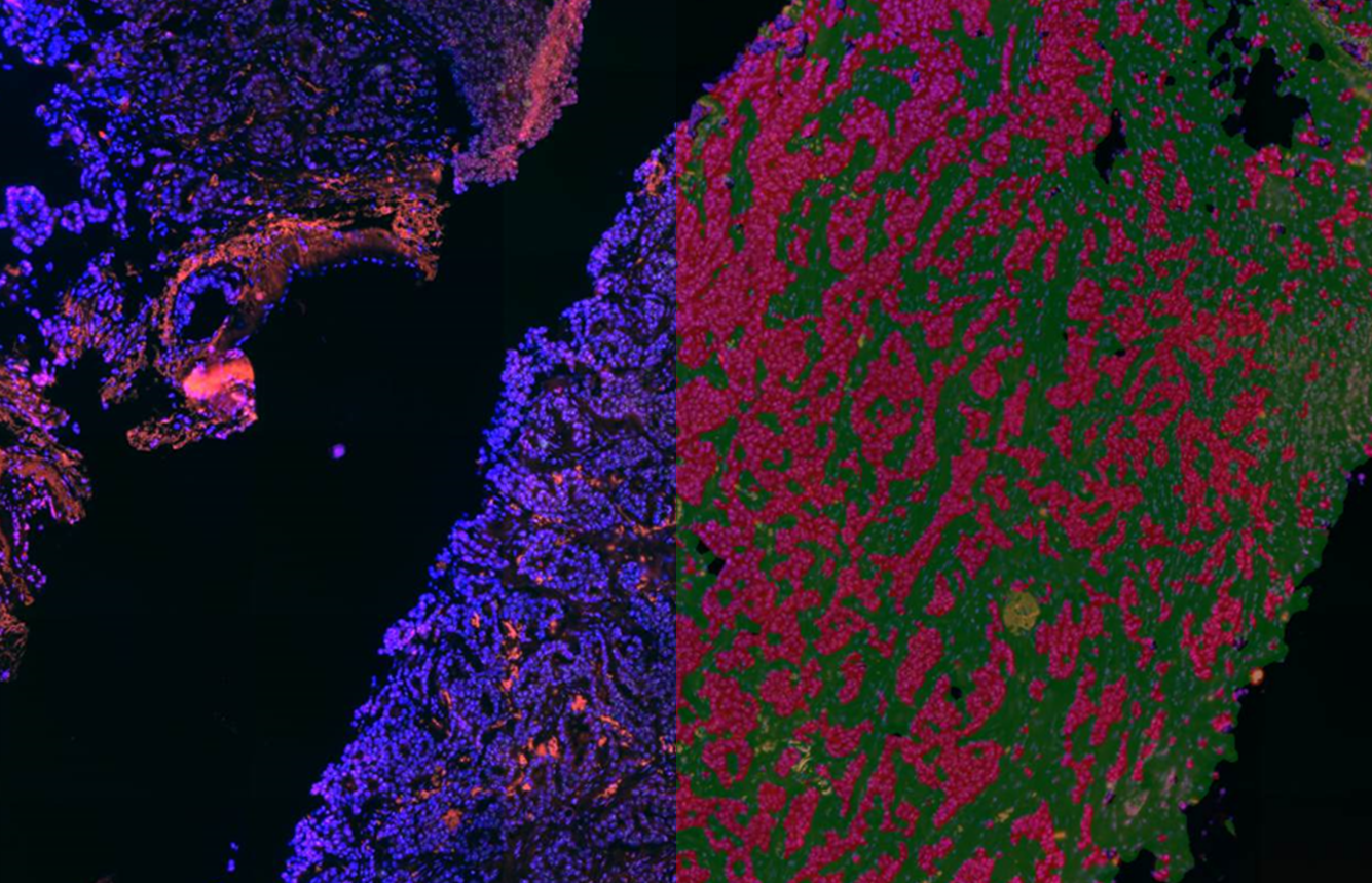
Estrogen stained infiltrating ductal carcinoma in breast tissue
by PatternQuant and NuclearQuant
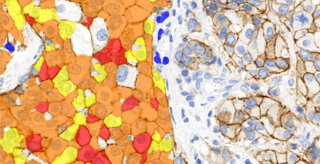
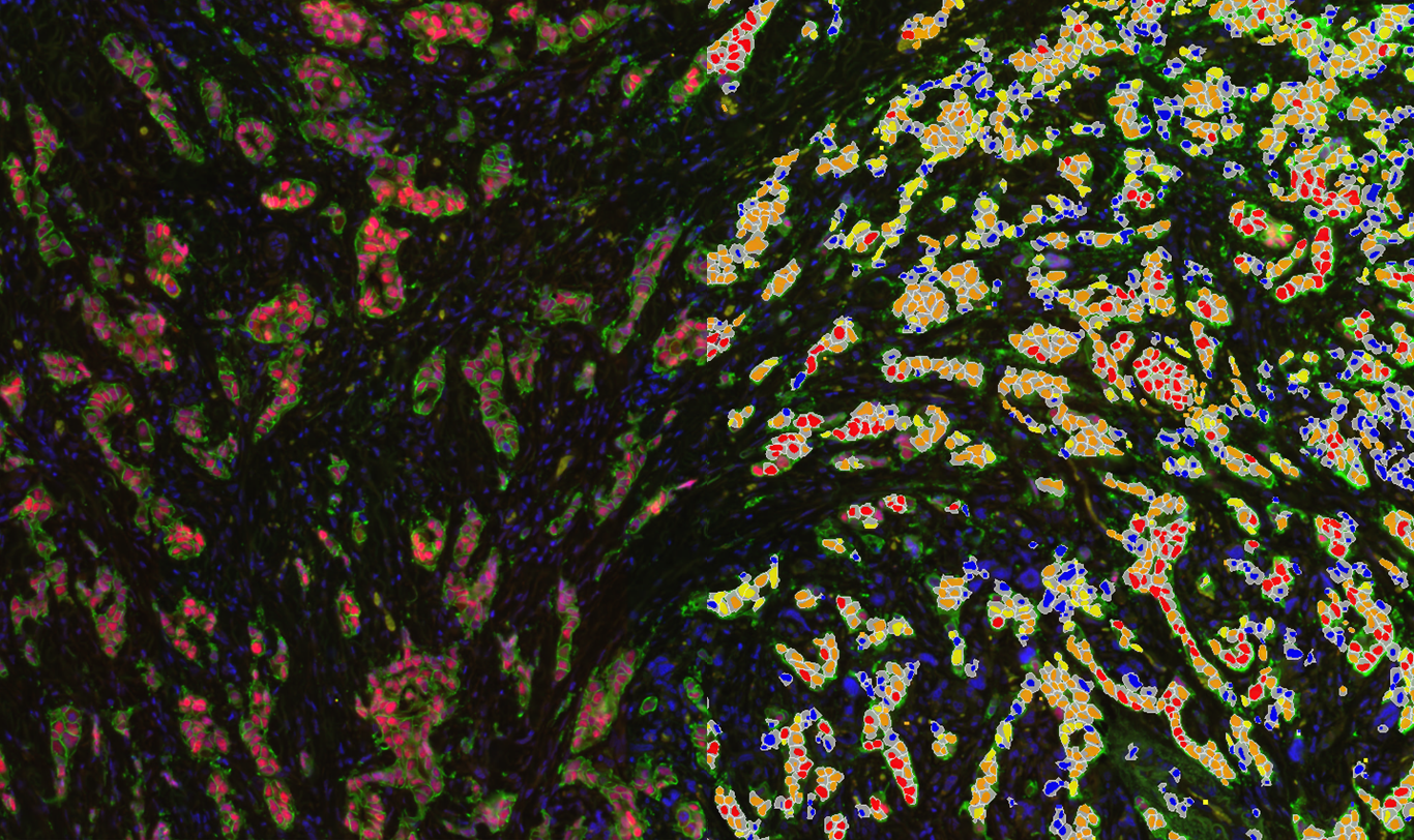
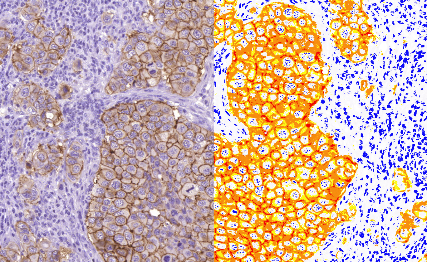
Estrogen-Her2 stained breast tissue analysis
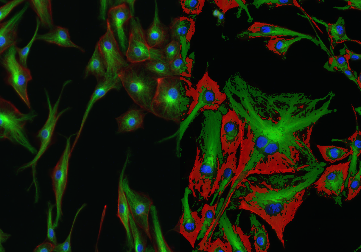
by CellQuant
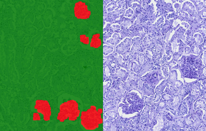
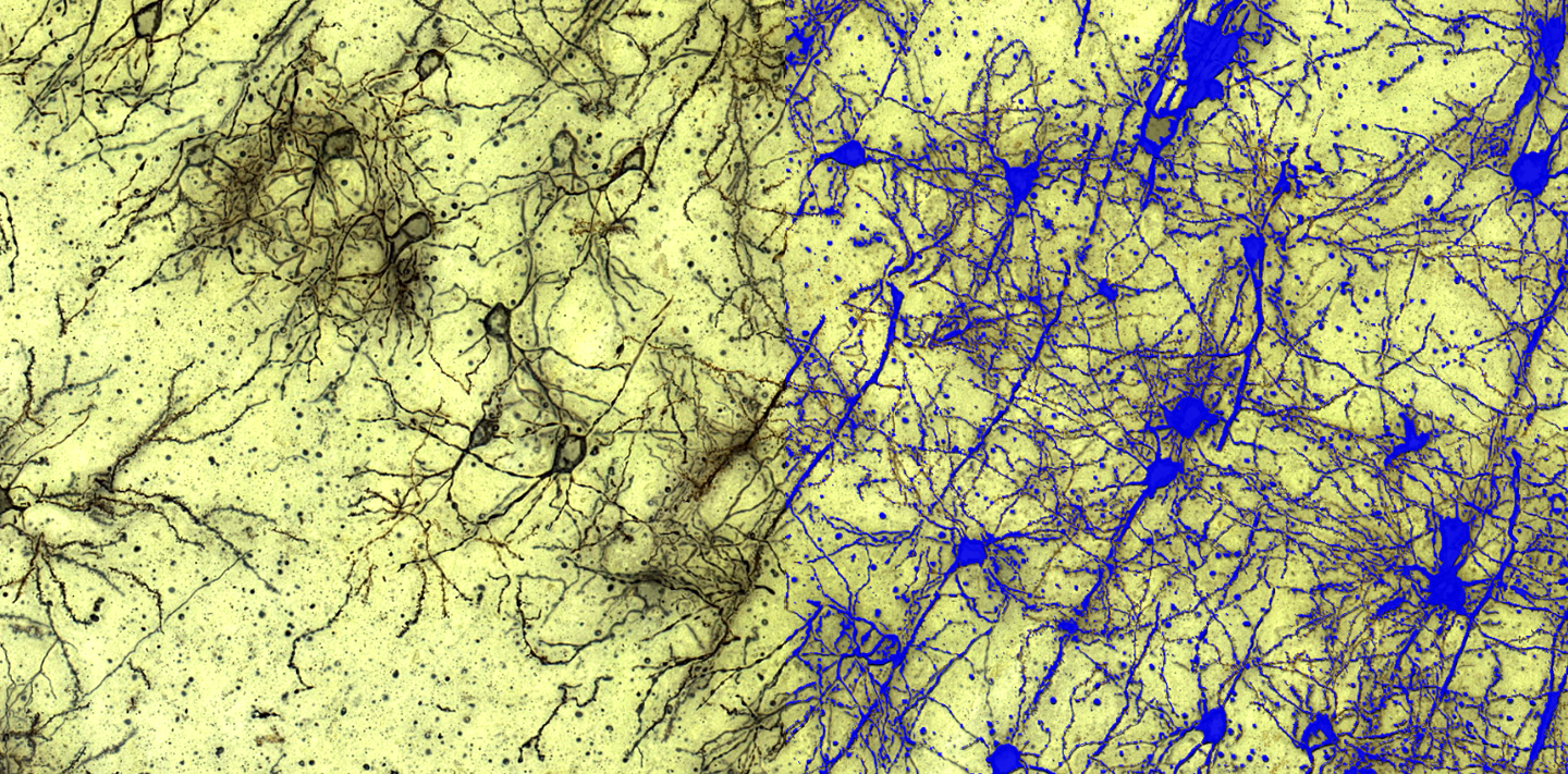
Golgi stained neuron network detection in the rat cortex
by HistoQuant
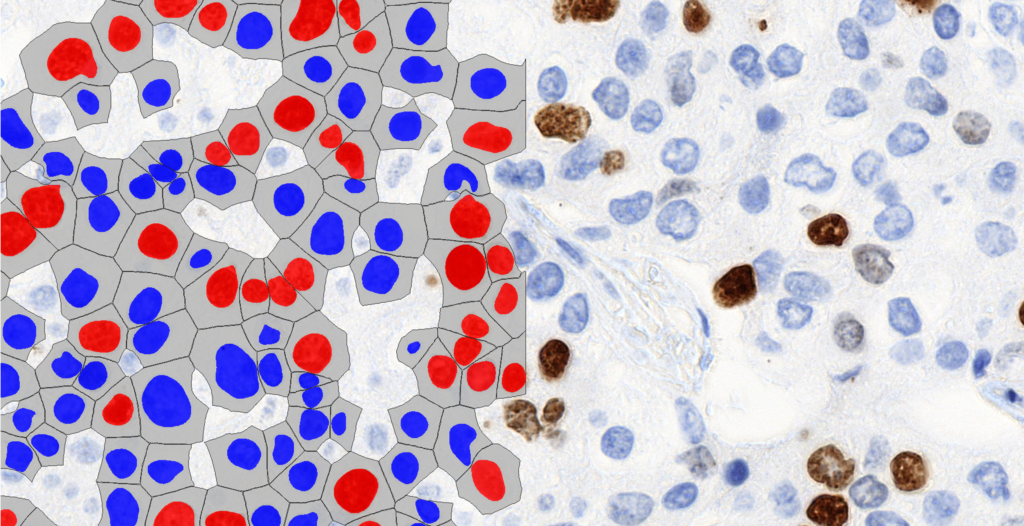
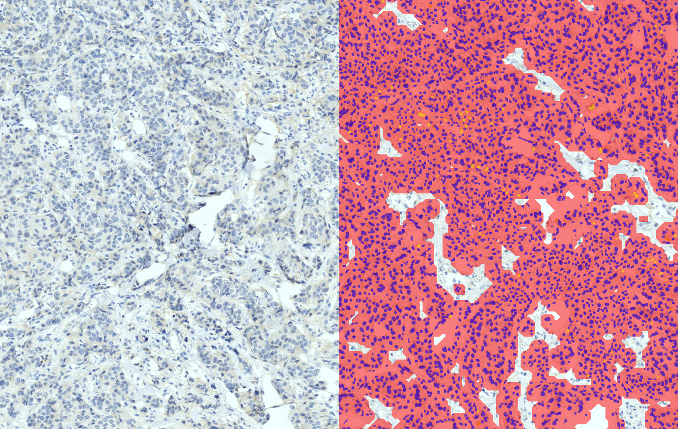
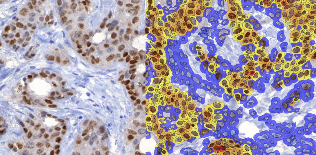
Nuclei classification in the tumour area of progresterone stained breast tissue
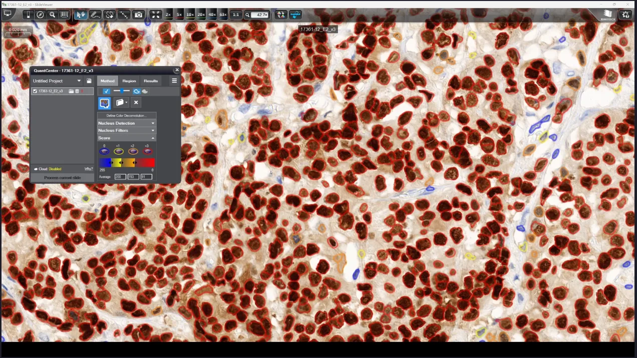
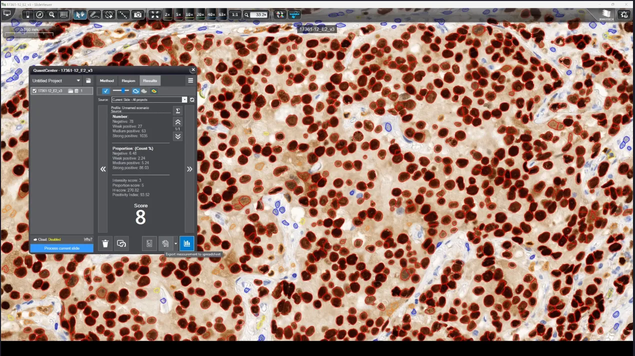
by NuclearQuant
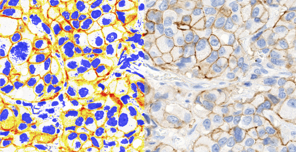
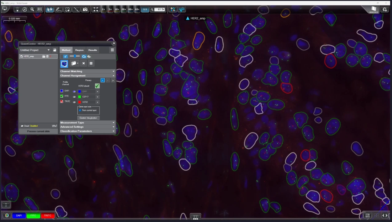
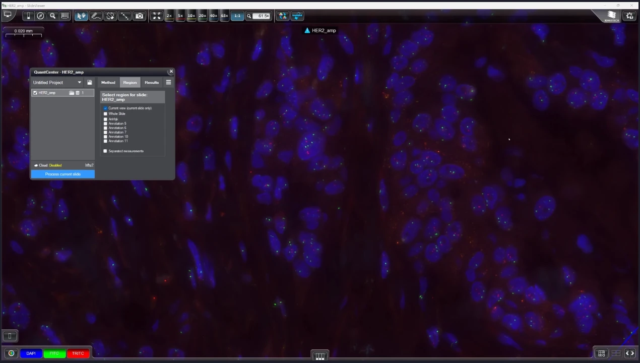
Her2 stained breast tissue analysis
by DensitoQuant
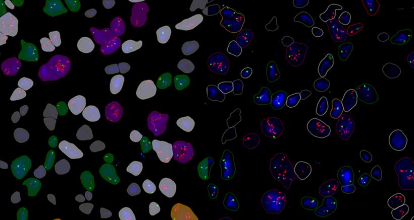
Her2-Cep17 DNA stained breast tissue classification
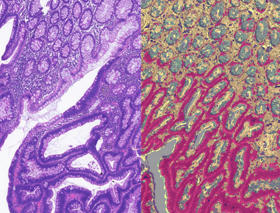
by HistoQuant
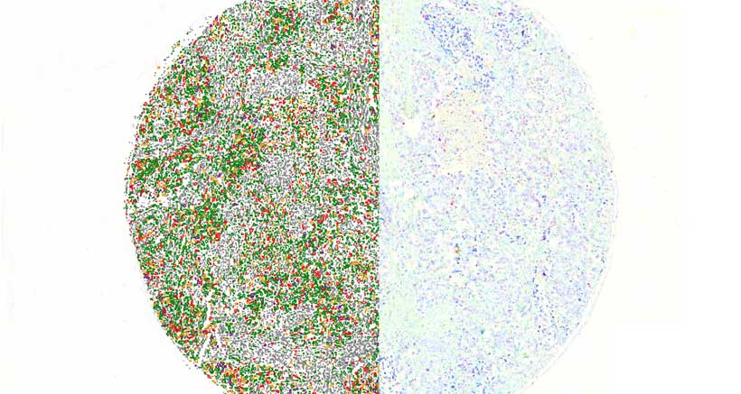
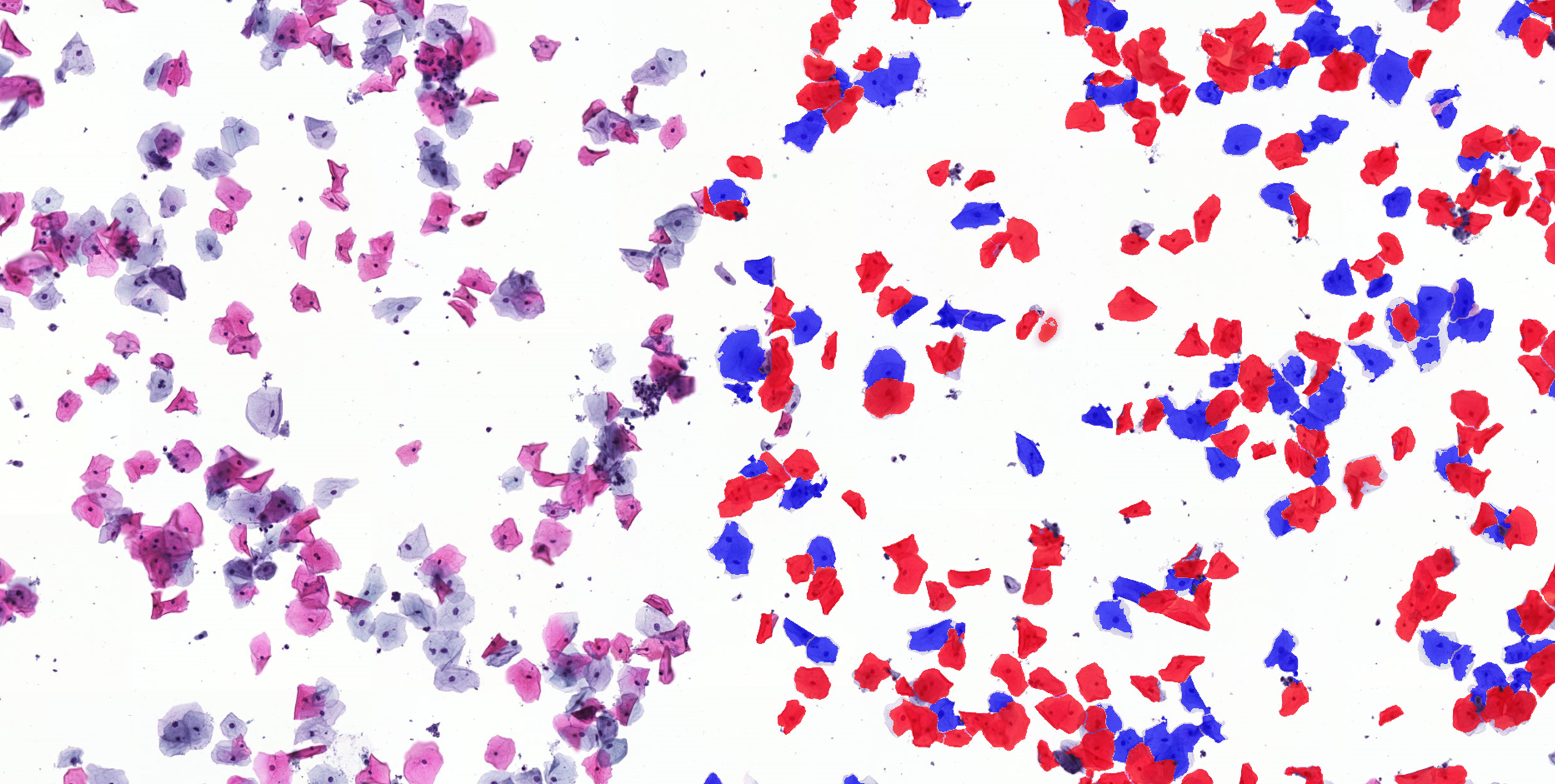
Nuclei classification on cytology smear sample
by HistoQuant
Progresterone stained cytoplasm classification
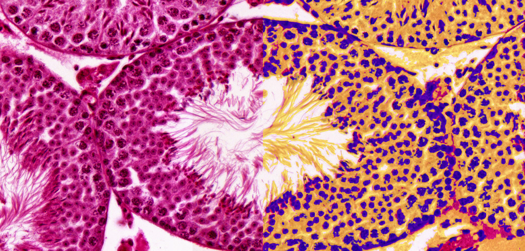
by CellQuant
Testicle organ with sperm cells analysis
by HistoQuant
QuantCenter
Key features
Modular and Customizable Image Analysis
Unlike traditional image analysis software, QuantCenter offers a modular framework, allowing users to select and combine specialized tools for precise whole-slide quantification.
Customizable AI-driven analysis enables adaptation to specific research needs, providing greater flexibility than static competitor solutions.
Comprehensive Range of Analysis Modules
Advanced segmentation and classification tools, including HistoQuant, PatternQuant, and PatternQuant Plus, ensure accurate stain identification and deep-learning-driven tissue recognition.
Targeted quantification with NuclearQuant, MembraneQuant, CellQuant, DensitoQuant, FISHQuant, and CISHQuant, covering all major staining techniques.
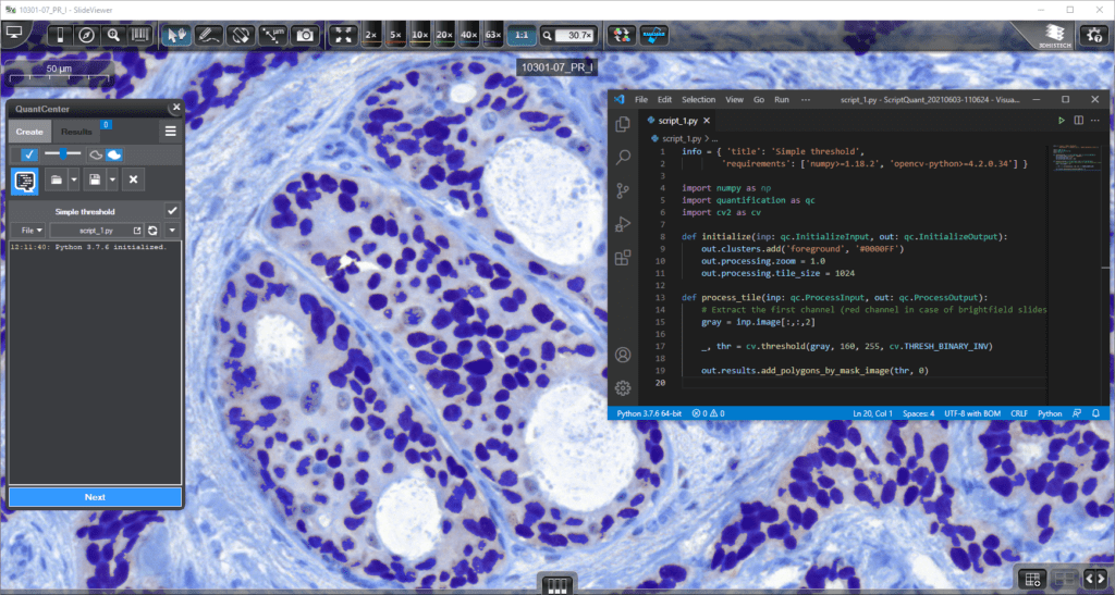
Custom script integration via ScriptQuant, offering unmatched flexibility in digital pathology.
Superior Performance and Batch Processing
BatchAnalysis-ProcessingQueue allows multiple digital slides to be processed simultaneously, increasing efficiency and throughput.
QuantServer ensures high-speed processing and centralized data storage, optimizing workflows for large-scale research studies.
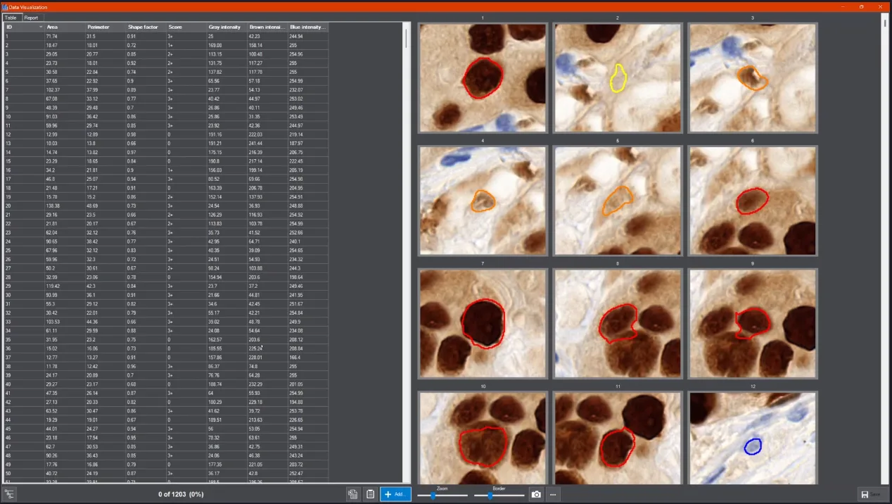
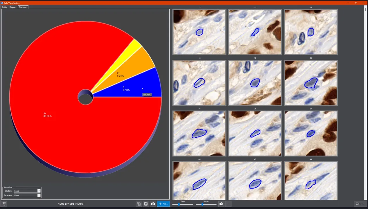
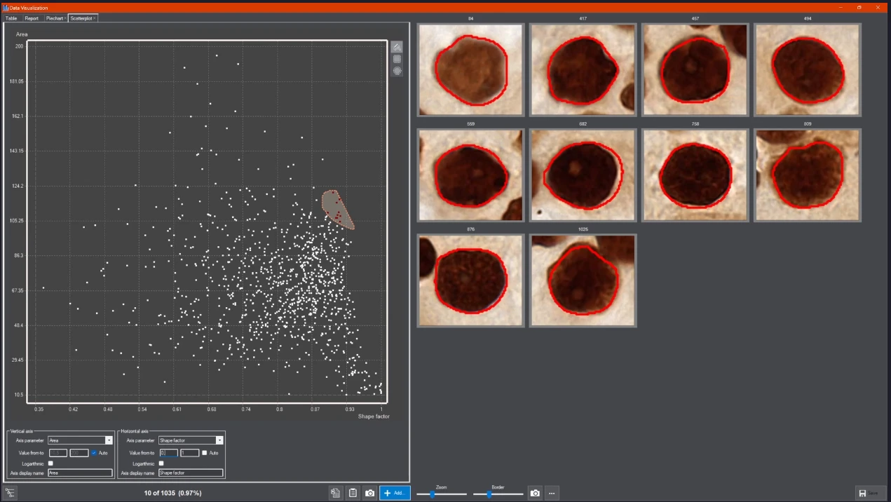
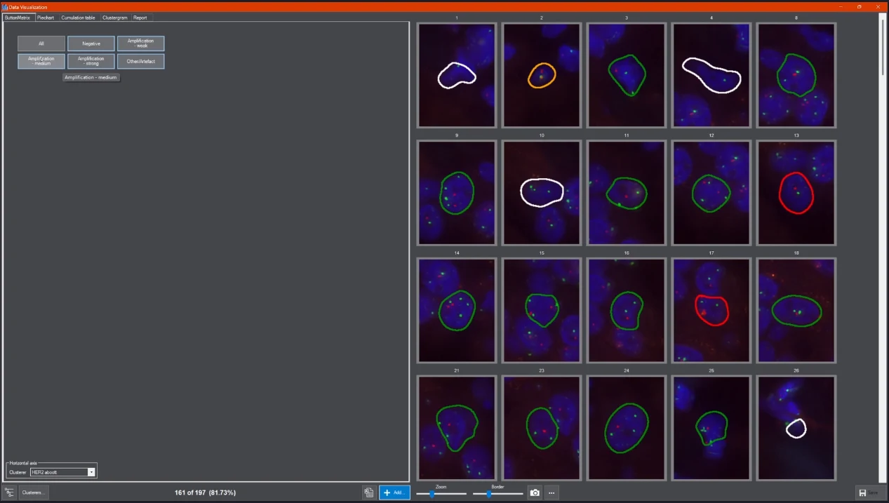
Advanced Data Visualization for Better Insights
Integrated Data Visualization (DVT) tools, including scatterplots, pie charts, histograms, and tables, enable intuitive and real-time result interpretation.
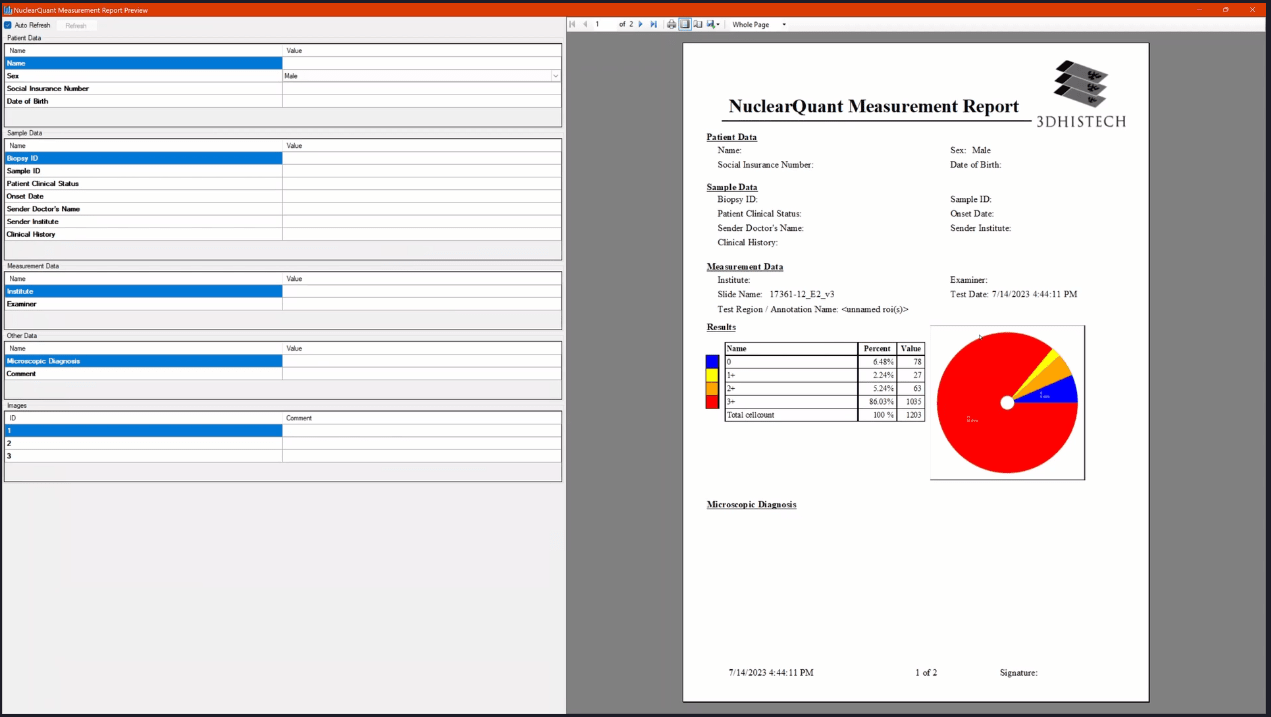
Quantitative analysis results can be easily exported for reporting and collaboration.
Seamless Integration with Digital Pathology Workflows
Fully compatible with SlideManager, ensuring smooth execution of analysis on study samples without requiring additional manual steps.
Supports a wide range of whole-slide image formats, making it a universal solution for diverse pathology laboratories.
Scalable for Research and Clinical Applications
Suitable for small-scale research and high-throughput clinical studies, with adaptable computational power and server-side processing.
Enables standardized, reproducible image quantification, essential for clinical diagnostics and drug development.