The Pannoramic Scanner Software streamlines digital pathology with automated workflows, high-precision scanning, and flexible imaging modes, making digital pathology faster, more reliable, and easier to manage. Users benefit from barcode recognition, Z-stack imaging, and real-time teleconsultation, reducing errors and increasing efficiency. It combines superior image quality, high “ TEMPLATES throughput, and intuitive operation, ensuring seamless integration into institutional, research and educational workflows.
Key features
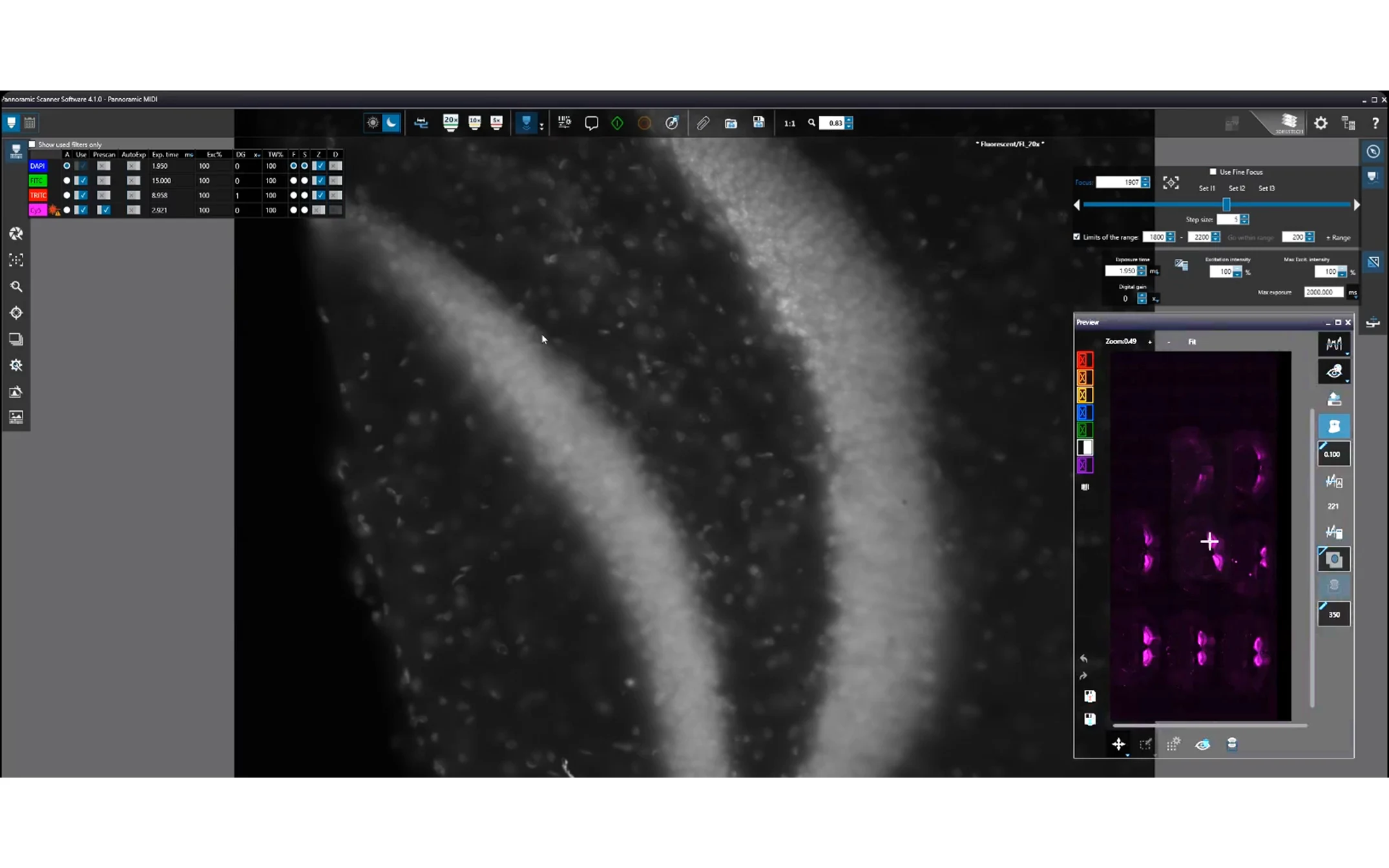
Advanced Whole Slide Imaging (WSI) for High-Quality Digital Pathology
Provides exceptional image resolution for brightfield and/or fluorescence scanning and enables smooth transition between imaging modes (all RX scanners support fluorescence except P1000 & P480). Ensures high-precision digitization of histology slides, reducing scanning errors and allowing for accurate research analysis.
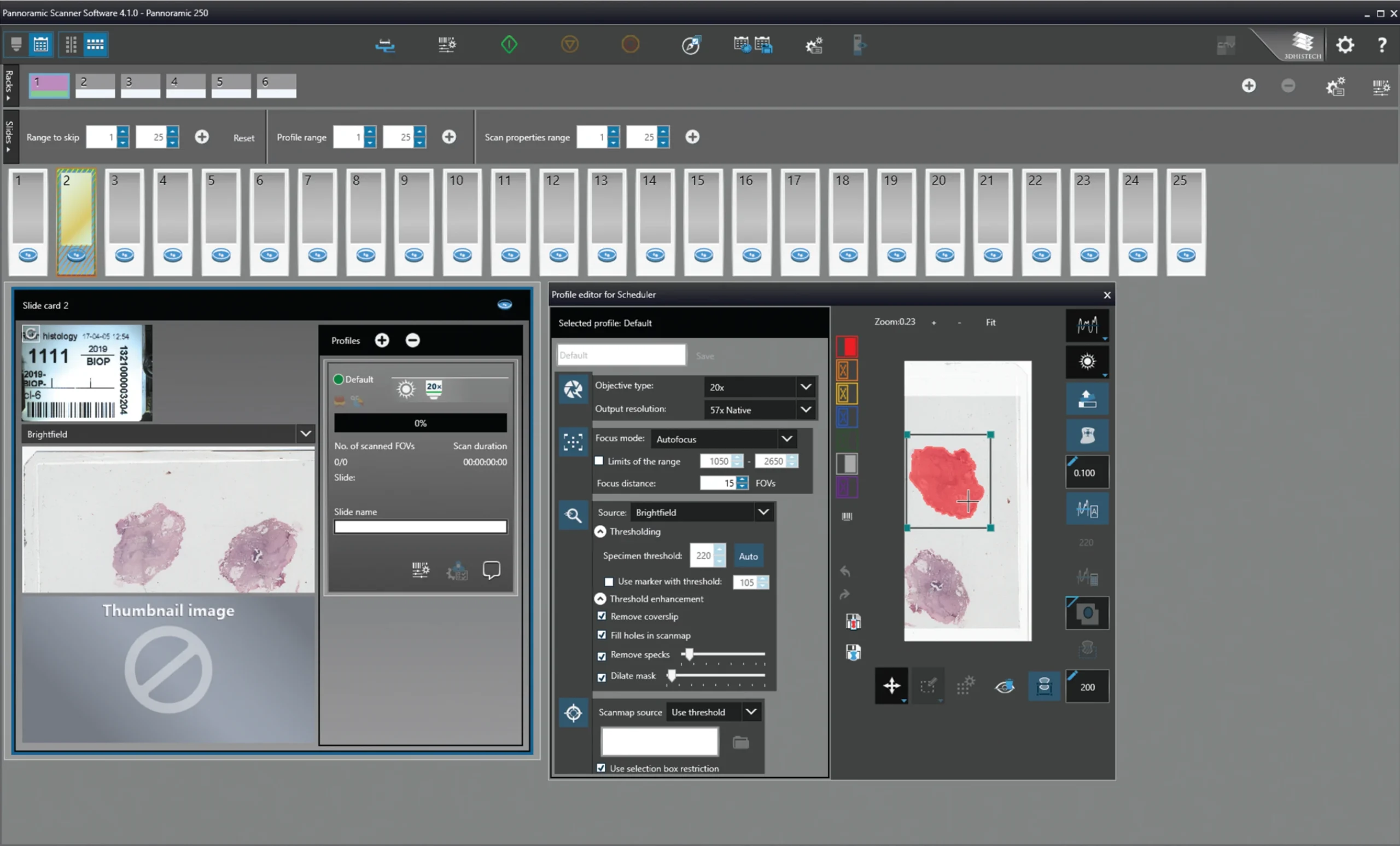
Automated and High-Speed Scanning
Fully automated scanning process with batch processing capabilities. Increases efficiency by reducing manual intervention, allowing researchers to focus on analysis rather than slide handling. Enhances throughput with optimized slide loading and scanning order, making it ideal for high-volume workflows.
Flexible Scanning Modes and Customization
Multiple scanning profiles, Z-stack imaging, and extended focus options. Enables customization based on specific research needs, improving flexibility. Allows for tailored scanning settings to match sample variability, reducing rescans and workflow interruptions.
Real-Time Teleconsultation & Image Sharing
Supports real-time remote access to digitized slides. Enables collaboration between researchers and pathologists from different locations. Facilitates rapid second opinions and remote slide analysis, accelerating research workflows.
AI-Based Tissue Detection and Barcode Recognition
Minimizes errors in slide tracking and positioning with built-in barcode recognition for automatic slide identification and AI-powered tissue detection. Ensures workflow standardization, reducing manual data entry and increasing reliability.
High-Resolution Image Management
Allows seamless integration with LIMS and pathology databases. Offers multiple image compression options and file formats (DICOM, non-overlapping MRXS). Reduces storage burden while maintaining high-quality imaging for easy sharing and archival.
High-Speed Fluorescence Imaging (All RX scanners except the P1000 & P480)
Provides superior fluorescence detection with motorized switching between brightfield and fluorescence. Supports up to 50 fluorescent channels with 9 physical filter positions. Enhances research workflows for multiplex imaging, critical for cancer and molecular pathology.
Seamless Integration with Research & Educational Platforms
Ensures smooth operation within modern lab environments. Compatible with Windows 11 and research-based workflows. Eliminates compatibility issues, enhancing productivity in pathology and life sciences research.
System requirements
Requires the All-in-one Server PC
Operating System: Microsoft Windows 11 Professional 64-bit EN.
Hardware Requirement:
– Processor: AMD Threadripper PRO 5975WX (32 Cores / 64 Threads) or similar.
– RAM: 128 GB DDR4.
Storage:
– 256 GB SSD (Primary Drive) – 2x 1 TB HDD (Secondary Storage)
Graphics:
– NVIDIA T400 or equivalent/higher
– Full HD (1920x1080p) display, 96 dpi resolution
Input Devices: Keyboard, Mouse.
Ports:
– 6x USB 2.0
– 4x USB 3.0
Ports & Connectivity:
– USB: Minimum 6x USB 2.0, 4x USB 3.0 (Pannoramic 250, 150, 75, MIDI III, Confocal, Desk II DW)
– USB 3.2 Gen 2 & USB-C Gen 2 support for high-speed data transfer (Pannoramic 1000, 480)
– CoaXPress (Pannoramic 1000, 480)
Additional Features:
– DVD SuperMulti SATA slim drive (Pannoramic 1000, 480)
– Support for external barcode reader integration
Software Features:
– Fluorescence Mode (not available for Pannoramic 1000 and 480)
– Extended Focus
– Z-Stack Imaging
– Barcode Reader (Licensed Feature)
– FRET Imaging Mode
Recommended Network Bandwidth
The recommended bandwidth for operating the Pannoramic Scanner Software effectively is:
– Minimum: 1 Gbps Ethernet (10/100/1000 Mbps)
– Recommended for High-Throughput Workflows: 10 Gbps Ethernet (RJ-45)
Ethernet:
– 10/100/1000 Mbps (Pannoramic 250, 150, 75, MIDI III, Confocal, Desk II DW)
– 10GbE RJ-45 (Pannoramic 1000, 480, 250)
For Large-Scale Multi-User Environments: Dedicated high-speed local network with low-latency connectivity:
– Ensures fast transmission of large whole slide image files without lag.
– Supports real-time teleconsultation and collaboration with minimal delays.
– Enables parallel scanning and image processing without network bottlenecks.
For best results, high-volume research and pathology labs should use 10 Gbps connections, especially when handling large datasets or using cloud-based image storage solutions.
