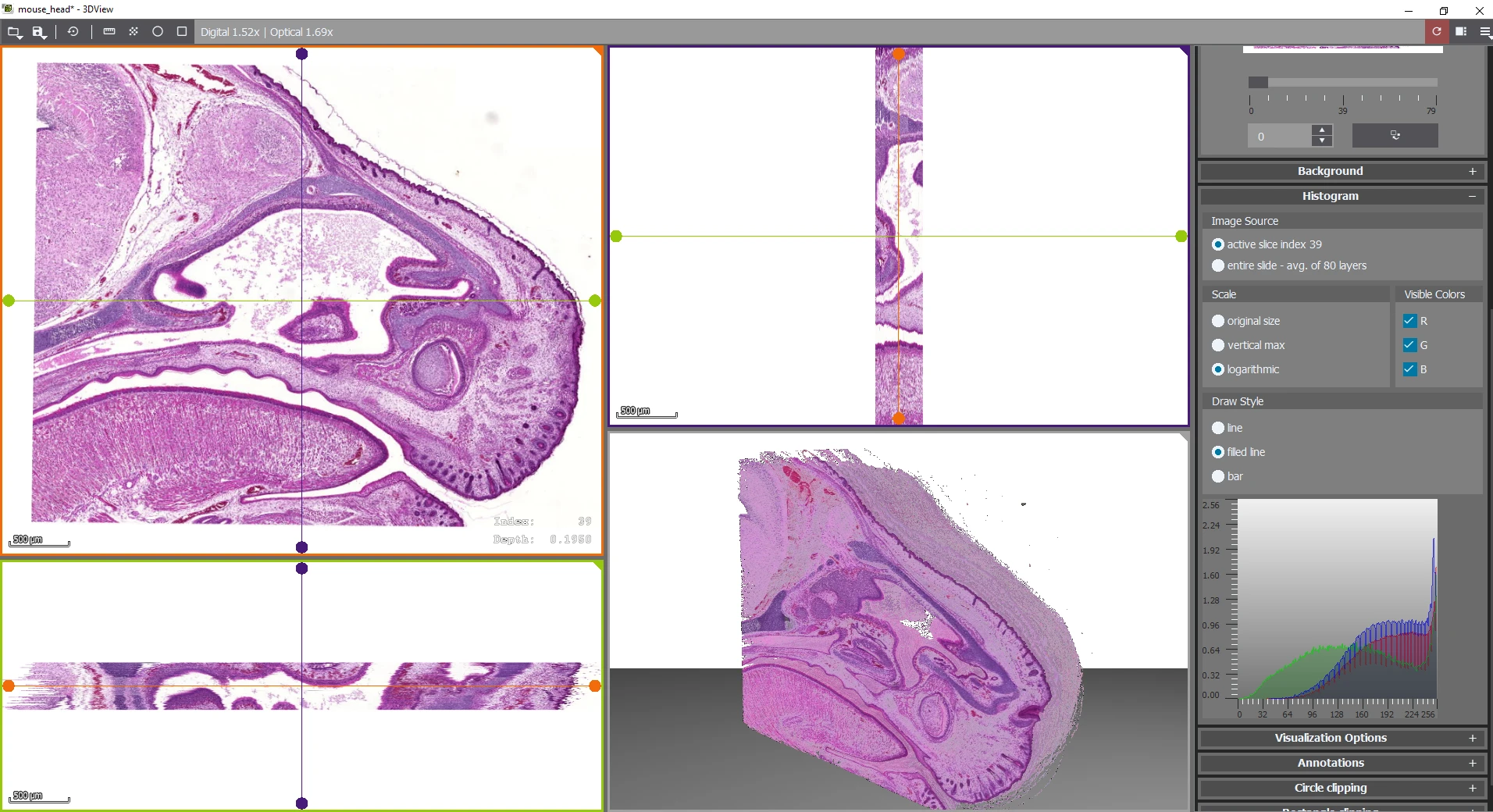
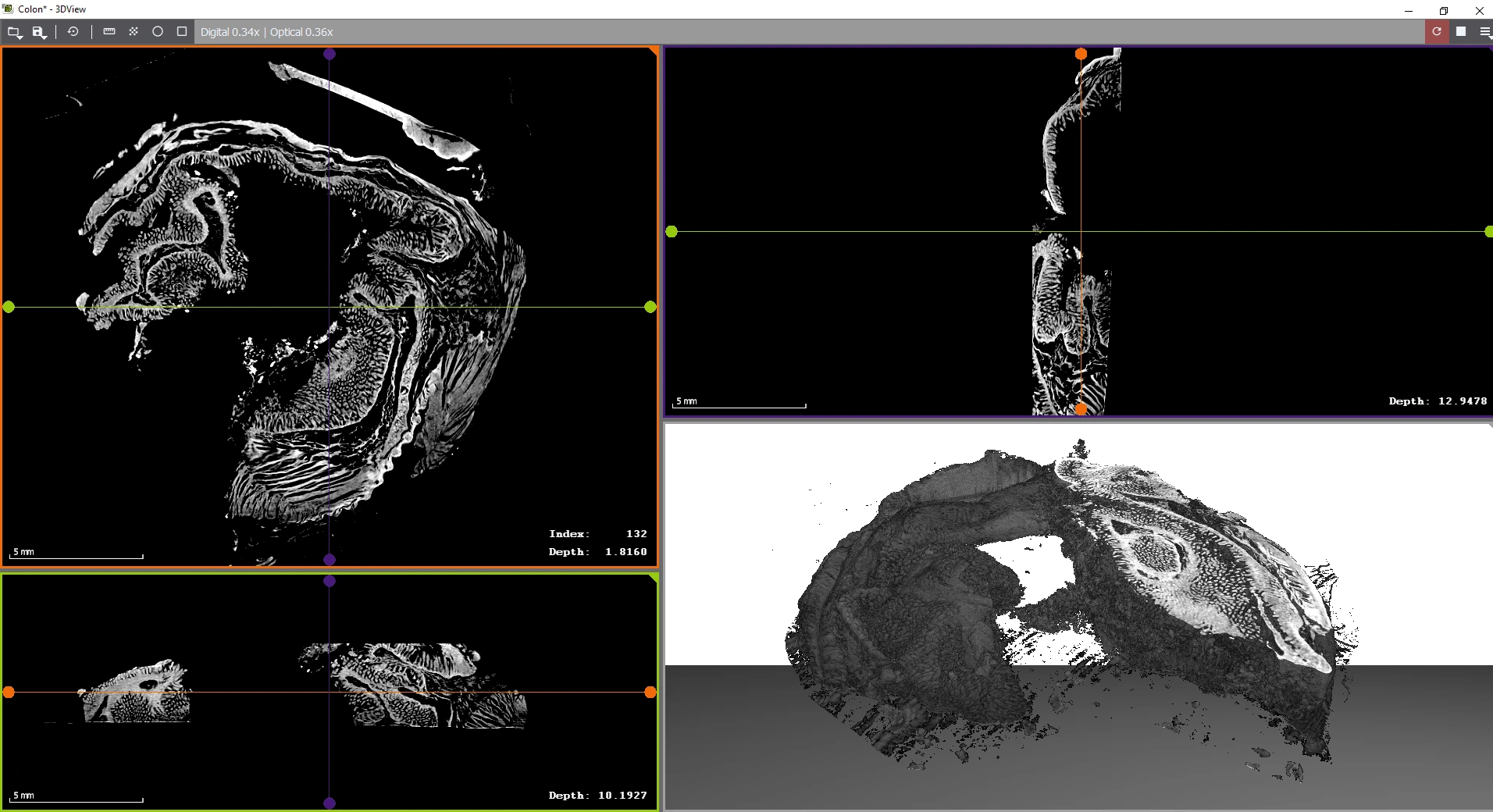
A powerful intuitive 3D imaging tool that goes beyond traditional microscope visualization. 3DView transforms 2D serial sections into interactive 3D reconstructions offering advanced visualization. It also supports micro CT images in VOL/VGI format for comprehensive tissue analysis.
Key features
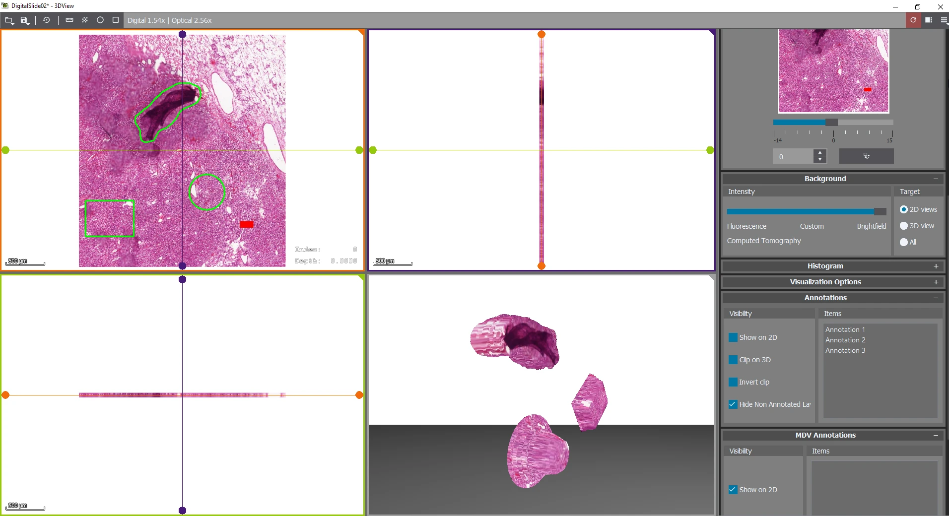
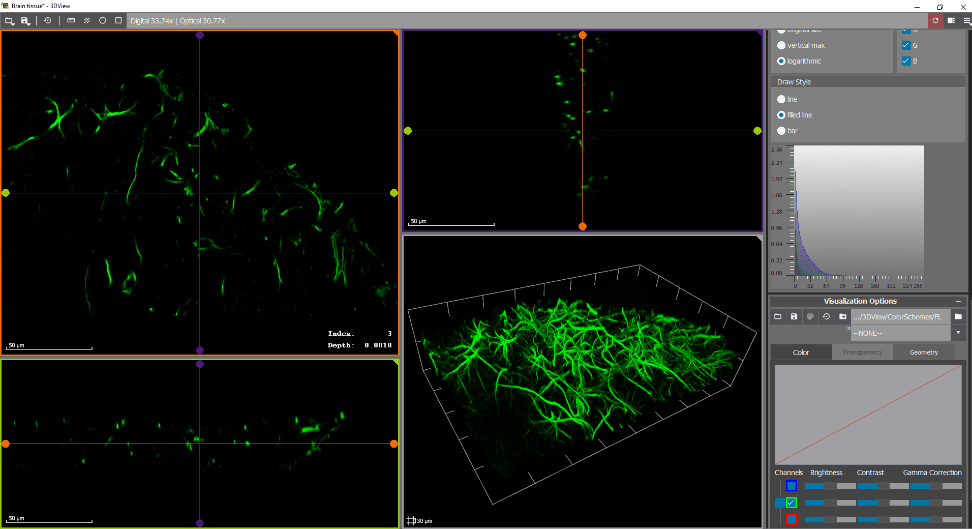
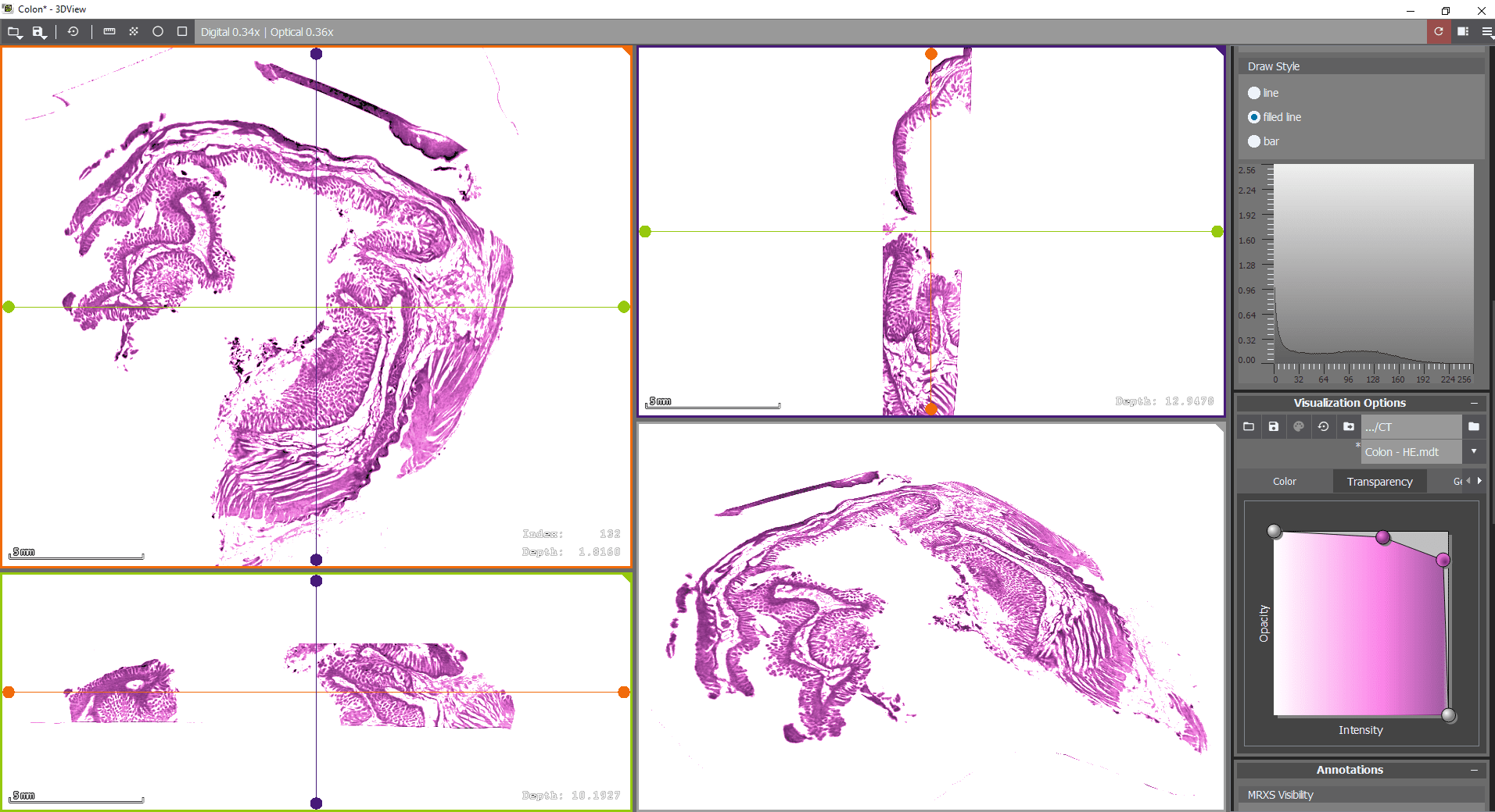
Advanced Visualization Options
Adjust brightness, contrast, gamma, and RGB channels select and edit multiple staining for optimal clarity.
Supports both 2D slice view (arbitrary sections) and 3D volume view for a
comprehensive understanding of tissue structures.
Creates 3D reconstructions from Z-stack slides, offering an enhanced perspective
beyond standard imaging.
Precision Measurement Tools
Accurately assess metric distances within the 3D model for more detailed analysis.
Flexible Export Options
Export reconstructions as 2D image series or full 3D volumes, with adjustable channel data, alignment, magnification, and format settings.
Generate high-resolution video exports and publication-ready snapshots for presentations, research, and documentation.
Industry-Leading Compatibility
Supports MRXS by 3DHISTECH, SVS by Leica Aperio, CZI by Zeiss, NDP by Hamamatsu, and VSI by Olympus, ensuring seamless integration with major whole slide imaging formats.
System requirements
– Operating system: Windows 7 SP1, 8. 8.1 or 10 (64-bit version)
– Graphics processing unit: OpenGL 3.3 capable (with dedicated video memory of 512 MB minimum, 1 GB recommended), such as from AMD/ATI, Barco, Intel, or NVIDIA
– CPU: Intel Core i7 or comparable
– RAM: 4 GB minimum, 8 GB recommended
