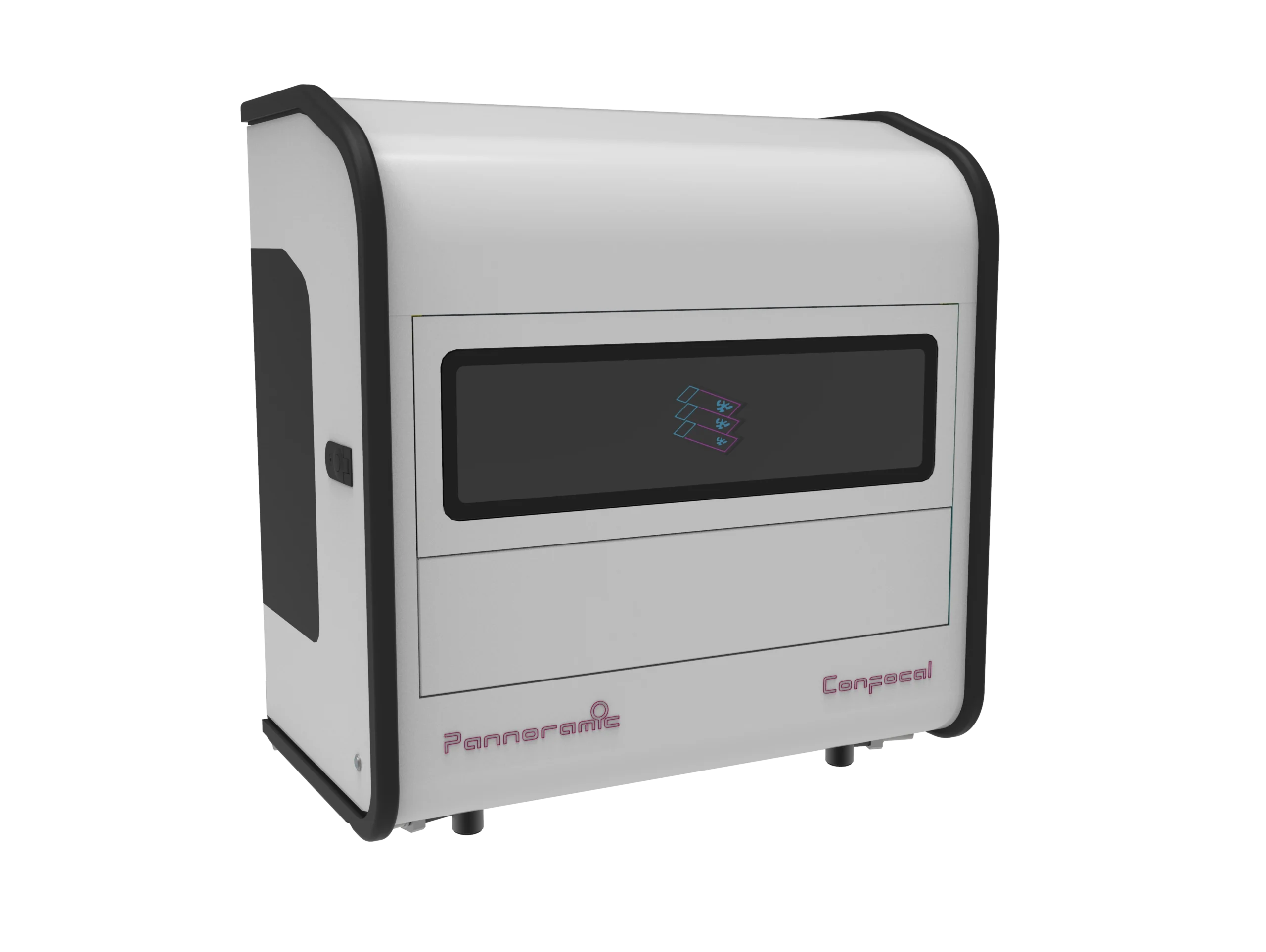
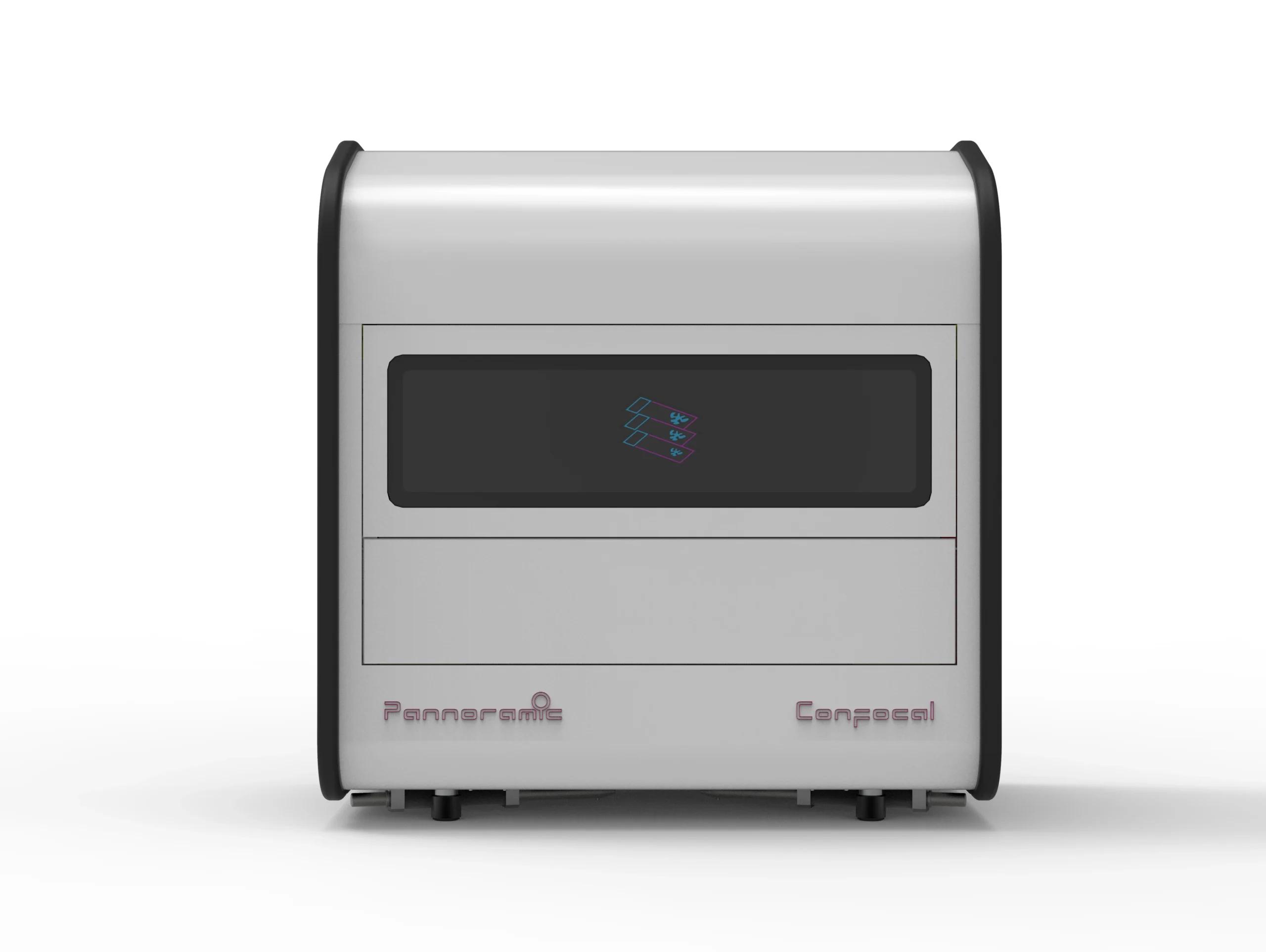
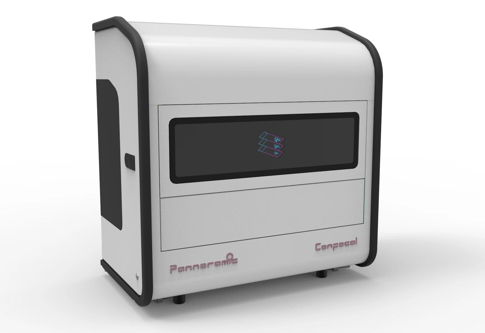
Pannoramic Confocal Digital Scanner
Advancing Digital Pathology for Research with the Pannoramic Confocal
The Pannoramic Confocal Digital Scanner represents a groundbreaking advancement in digital pathology and fluorescence imaging, integrating confocal microscopy with whole-slide imaging technology to achieve unparalleled clarity, depth, and contrast. Unlike traditional WSI scanners, which capture flat, two-dimensional images, the Pannoramic Confocal excels in Z-stack scanning, generating detailed, multi-layered 3D reconstructions of tissue samples.
This advanced system utilizes structured illumination confocal imaging, overcoming the limitations of spinning pinhole-disc techniques to maximize light efficiency, minimize photobleaching, and enhance signal-to-noise ratios. Its high-speed scanning capabilities – up to 20x faster than conventional confocal systems – ensure optimal performance in multiplex imaging, fluorescence-based research, and molecular pathology applications.
With its automated water immersion system, scientific CMOS camera for ultra-low-noise imaging, and AI-driven digital differential interference contrast (SW DDIC) for brightfield visualization, the Pannoramic Confocal sets itself apart for research pathology, drug development, and translational research.
Key Features:
Confocal Z-Stack Scanning for 3D imaging
- Multilayer (Z-stack) scanning with up to 100 optical sections
- 3D tissue reconstruction for in-depth morphology studies
- Eliminates out-of-focus blur, enhancing depth and contrast
Unmatched Speed and High-Resolution Imaging
- Scanning speed up to 20x faster than conventional confocal systems
- SCMOS camera with low-noise (1.3 e-) imaging ensures high sensitivity
- Resolution down to 0.10 µm/pixel, supporting superior image clarity
Innovative Structured Illumination Technology
- Overcomes limitations of spinning-disc confocal microscopy
- Minimizes photobleaching while maximizing light collection
- Colocalized imaging available in fluorescent and brightfield modes
Advanced Fluorescence and Brightfield Imaging
- 6-channel solid-state fluorescence light engine (15,000-hour lifespan)
- Supports quad-band and multi-channel fluorescence imaging
- Brightfield imaging enhanced by SW DDIC technology
Automated Water Immersion and Anti-Bleaching Solutions
- Water immersion system for high NA objectives, improving signal intensity
- Minimized sample bleaching through precise light control & LED-based excitation
- LED light engine lifespan exceeds 15,000 hours, reducing long-term costs
Optimized Image Handling and Data Export
- Multiple export formats including JPG, TIFF, and lossless 3D imaging exports
- Seamless integration with third-party analysis software
- Capacity: 11+1 slides
- Compatible Slide Formats: 25.5 mm x 75.5 mm, 1 mm thickness
- Dual-objective support with motorized objective changer.
- Default Objectives:
- Zeiss Plan-Apochromat 20x/0.8 NA
- Zeiss C-Apochromat (W) 40x/1.2 NA (water immersion)
- Camera Type:
- 5 Mpx, 16-bit, scientific CMOS camera (low noise: 1.3 e-)
- Resolution:
- Brightfield: 0.4 µm FWHM (40x 1.2 NA objective)
- Confocal Sectioning: 1.43 µm FWHM (40x 1.2 NA objective)
- Speed:
- Brightfield Scanning Speed: 13 minutes for a 15 mm x 15 mm sample at 0.325 μm/pixel.
- Fluorescence Scanning Speed: 30 minutes for a 15 mm x 15 mm sample with 3 fluorescent channels at 20 ms exposure per channel.
Fluorescent Illumination:
- 6-channel solid-state light engine (15,000-hour lifetime)
- Supports DAPI, FITC, TRITC, Cy5, and custom filter sets
Brightfield Illumination:
- 3CCD-equivalent separated RGB LED
- Automatic threshold tissue detection using a USB preview camera
- Multilayer Z-stack scanning with adjustable layer distances (0.2–2 µm)
- Supports 1D and 2D barcode formats, including Code39, Code128, QR, and Data Matrix.
- Digital Slide Format: Proprietary MRXS (lossless or JPEG2000 encoding)
- Export Formats: Multi-channel or single-channel TIFF, JPEG, PNG, BMP
- 3D Image Export: Compatible with third-party 3D analysis software
- Dimensions (W x D x H): 97 cm x 58 cm x 103 cm (39” x 23” x 41”)
- Weight: 100 kg