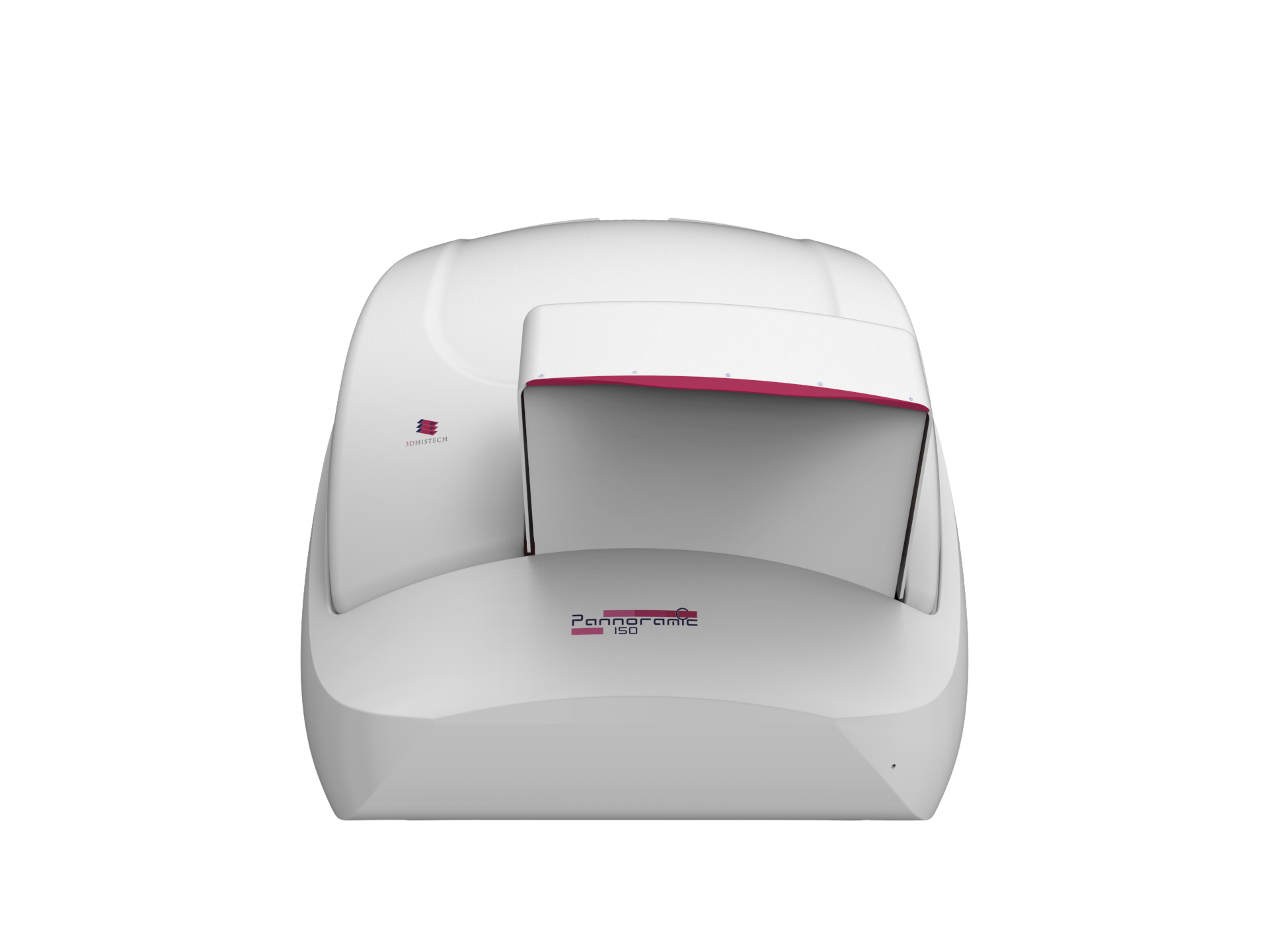
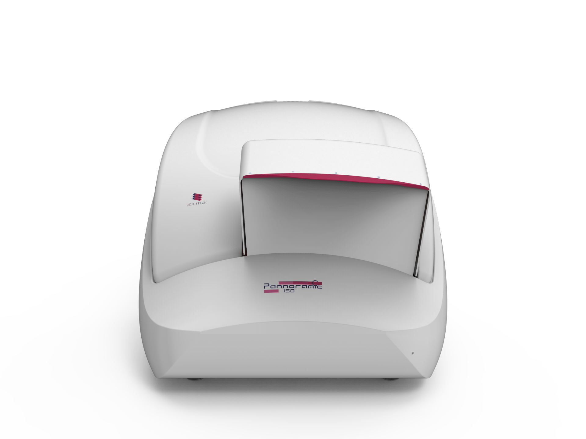
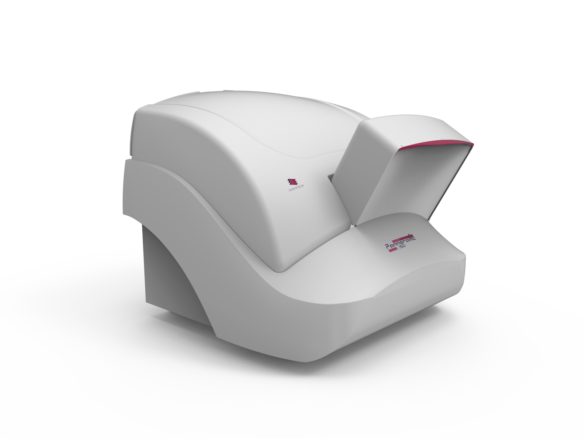
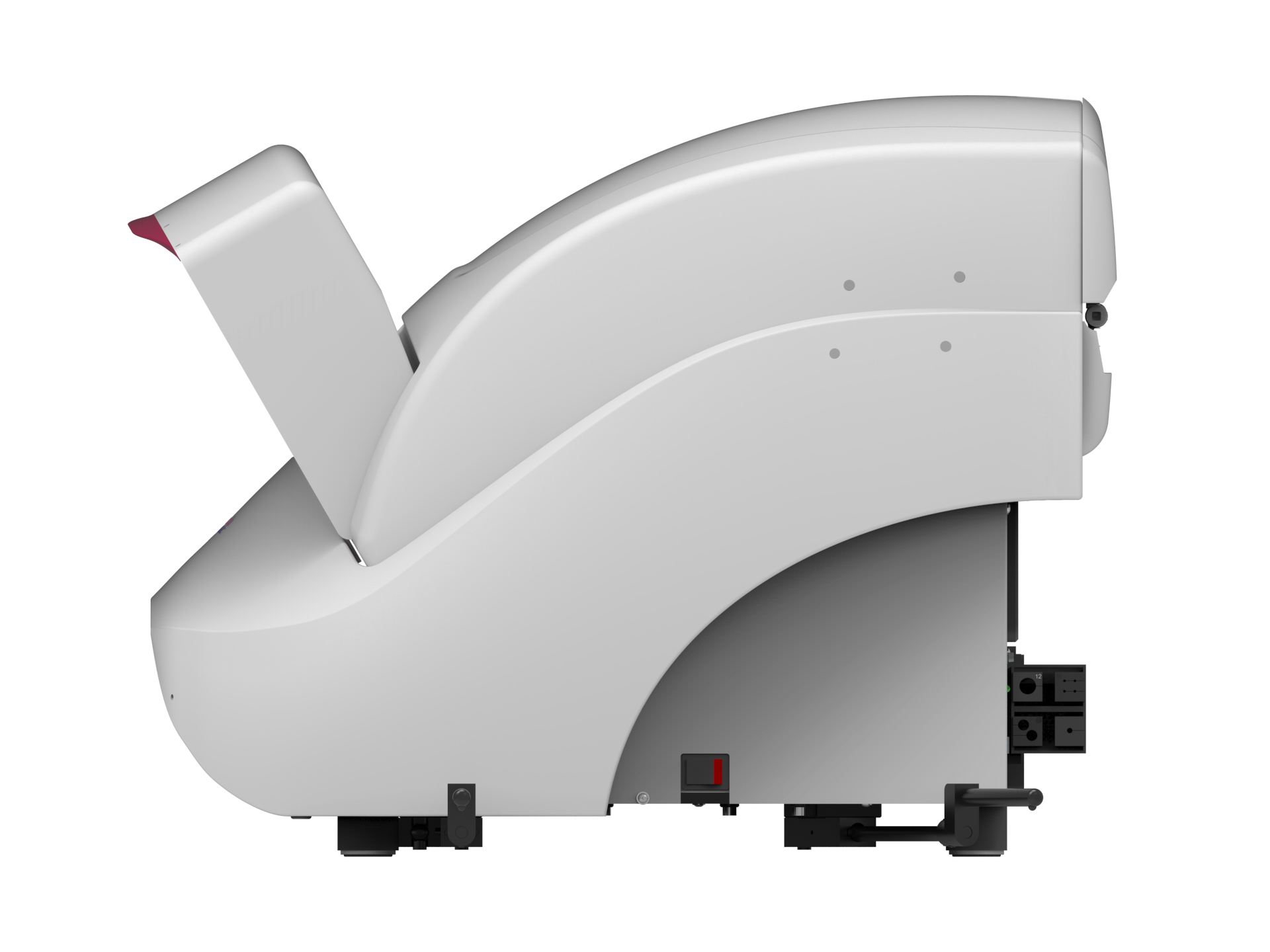
Pannoramic 150 Digital Scanner
Pannoramic 150 – High Throughput Digital Slide Scanner for Advanced Brightfield and Fluorescence Imaging
The Pannoramic 150 sets a new standard in digital pathology and whole-slide imaging with high-speed, high-resolution scanning for both brightfield and fluorescence applications. Designed for high-throughput research, it combines cutting-edge optics, automation, and software integration to deliver unparalleled performance, accuracy, and efficiency. With a 150-slide capacity and continuous loading, it eliminates workflow bottlenecks, enabling labs to process more slides in less time. Its Plan-Apochromat 40x/0.95 NA objective achieves 103x magnification at 0.10 µm/pixel resolution, producing publication-quality brightfield images with exceptional clarity. The continuously moving stage and 72 fps brightfield color camera further enhance speed without sacrificing detail.
A key differentiator is its exclusive 10x fluorescence objective, an innovation unique to 3DHISTECH that reduces fluorescence scan time by 50% compared to conventional scanners. Unlike competitor systems that rely solely on higher magnifications, the Pannoramic 150 combines high-speed fluorescence imaging with uncompromised resolution and sensitivity. This groundbreaking feature, paired with a back-illuminated 16-bit 4.2 MP sCMOS camera, 9-position motorized fluorescence filter changer, and high-end multiband light engine, provides unmatched speed and efficiency for multiplexed fluorescence imaging. Automated slide handling, barcode reading, and tissue detection further streamline the workflow, reducing manual workload and increasing productivity.
With faster, more reliable results, the Pannoramic 150 allows researchers to focus on data interpretation instead of troubleshooting technical issues. Less hands-on time means more productivity, as automated slide loading, barcode detection, and intelligent focus adjustment free up valuable research hours. The high-resolution brightfield and fluorescence imaging enhance data accuracy, delivering clearer, more detailed results for confident decision-making. Scalable for any lab size, it seamlessly adapts to both small-scale studies and high-volume research projects, ensuring efficiency without compromise. With its cutting-edge technology, automation-driven workflow, and superior imaging capabilities, the Pannoramic 150 is more than just a scanner, it’s an essential tool that simplifies your work, boosts efficiency, and improves research outcomes.
Key Features:
High-Throughput Scanning with 150-Slide Capacity – More Work Done in Less Time
Eliminates workflow bottlenecks and maximizes lab efficiency, enabling researchers to process more samples faster.
- The 150-slide capacity (6 magazines of 25 slides) with continuous loading allows you to scan large batches without manual intervention.
- Fully automated slide handling, barcode reading, and tissue detection reduce hands-on time, allowing you to focus on analysis rather than slide management.
Superior Brightfield Imaging – Exceptional Image Clarity for Confident Analysis
Produces publication-quality images for precise morphological assessment, reducing the need for rescanning.
- The continuously moving stage ensures smooth and high-speed scanning without compromising quality.
- A dedicated 5 MP color CMOS camera (72 fps frame rate) captures sharp, vibrant images with high color fidelity.
- Plan-Apochromat 40x/0.95 NA objective achieves 103x magnification at 0.10 µm/pixel resolution, making even the smallest histological details visible.
Twice the Fluorescence Scanning Speed – Faster, More Efficient Multiplexing
Speeds up fluorescence-based research, allowing for faster data collection, reducing sample exposure, and improving experiment efficiency.
- Equipped with a 10x fluorescence objective, cutting fluorescence scan time in half compared to conventional scanners.
- Back-illuminated 16-bit 4.2 MP sCMOS camera ensures optimal sensitivity, even for low-intensity signals.
- 9-position motorized fluorescence filter changer with multiband filter support enables highly flexible and multiplexed imaging.
Intelligent Autofocus and Imaging Enhancements – Less Manual Effort, Better Results
Minimizes manual adjustments, leading to more consistent and reliable results while reducing user fatigue.
- Fast multicolor fluorescence pre-scan for precise sample detection.
- Software-driven flat-field correction and deconvolution ensure uniform illumination and enhanced signal-to-noise ratio.
- Automated tissue and coverslip detection improves scanning accuracy.
Optimized Workflow & Connectivity – Seamless Integration with Your Lab Environment
Simplifies data management, collaboration, and remote analysis, saving time and making research workflows smoother.
- Fully automated operation with server-side barcode reading for efficient slide tracking and data integrity.
- Integration with Pannoramic Scanner Software, SlideViewer, SlideManager, and Quant for streamlined data analysis.
- Multiple scanning profiles and adjustable focus parameters adapt to a variety of research needs.
150 slides (6 magazines, 25 slides each) with automatic loading and continuous scanning
Dual-mode scanning (brightfield and fluorescence) with area scanning and autofocus
Included, for seamless transitions between BF and FL imaging modes without manual intervention
Included when two objectives are selected (supports two objectives)
1 minute 30 seconds for a 15 mm x 15 mm area at 0.19 µm/pixel resolution
8 minutes 30 seconds for a 15 mm x 15 mm area with 3 fluorescent channels at 20 ms exposure time
Optimized for high-throughput workflows with fully automated slide handling
Plan-Apochromat 10x/0.3 NA, 20x/0.8 NA, and 40x Corr/0.95 NA objectives
Up to 103x
Up to 98x (with 1.6x adapter)
MRXS format with export options to JPG, JPEGXR, BMP, PNG, TIFF, Roche TIFF, SVS, and DICOM
Slides measuring 75.0-76.2 mm (L) x 25.0-26.0 mm (W) x 0.9-1.2 mm (T) with No. 1 or 1.5 coverslips (0.13-0.19 mm)
Not explicitly specified
Down to 0.10 µm/pixel (with 40x objective)
Down to 0.10 µm/pixel (with 40x objective and 1.6x adapter)
LED-based transmitted light with 3DHISTECH LED FLASH technology (average lifetime ~60,000 hours)
Lumencor SOLA, Spectra III, or CoolLED pE-800 Light Engine with 8 solid-state sources (LEDs and lasers, 380-740 nm)
Automatic threshold tissue detection with a USB preview camera
75.0-76.2 mm (L) x 25.0-26.0 mm (W) x 0.9-1.2 mm (T)
Optional Z-stack (multilayer) scanning with 1-30 layers and adjustable distances (0.2-2 µm)
Supports 1D and 2D barcode types, including Code39, Code128, QR, and Data Matrix
Approx. 673 mm x 689 mm x 546 mm (scanner unit)
Approx. 49 kg (scanner unit)
Pannoramic 150 Digital Scanner
Specifications
Customizable Configurations
Available Objectives:
- 20x/0.8 NA (Default)
- 20x/0.8 NA and 40x/0.95 NA
- 10x/0.3 Plan-Apochromat objective (optional, available for fluorescence only)
Additional Options:
- Motorized Objective Changer (Included when two objectives are selected, as the standard configuration includes a single objective.)
- Multilayer (Z-Stack) Scanning
- Extended Focus Scanning