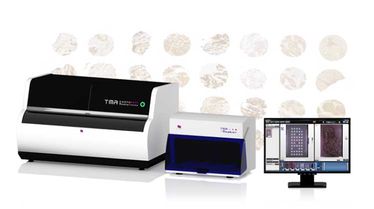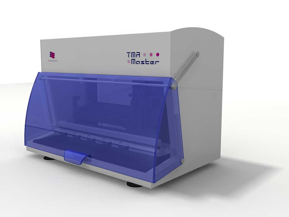
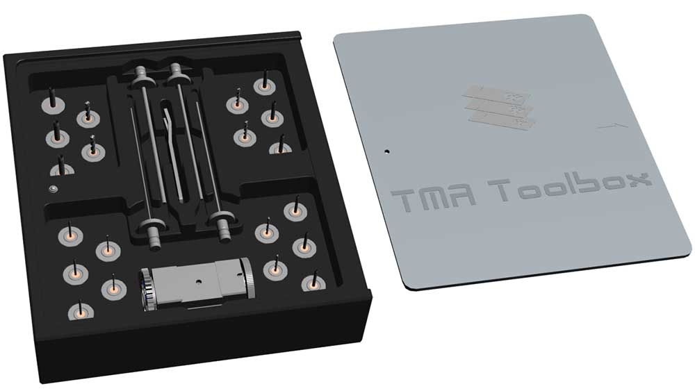
TMA Master II is the smallest fully automated tissue microarrayer on the market, it easily fits any laboratory bench.
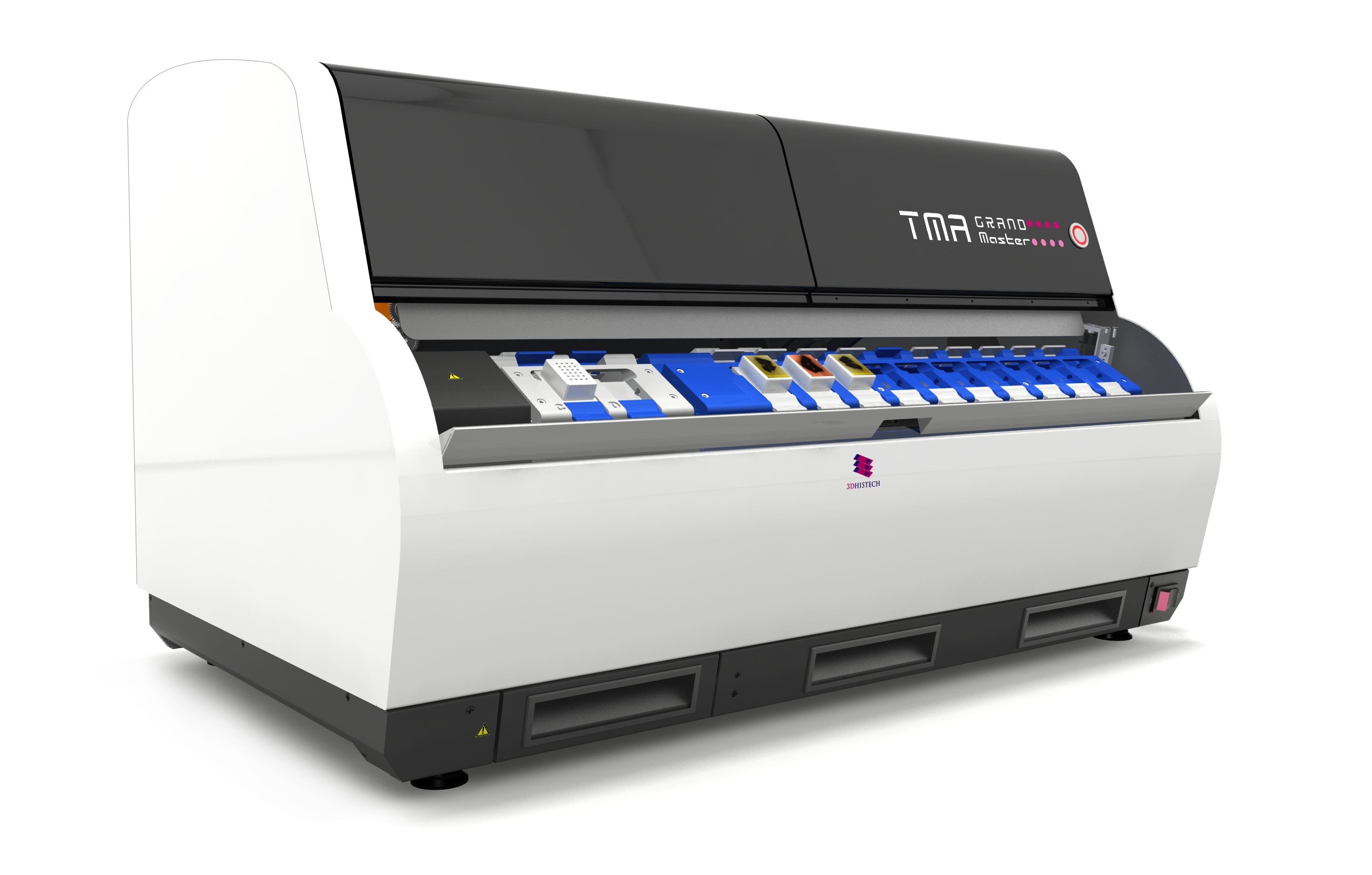
As the latest member of 3DHISTECH’s TMA product family, this device is based on our well-known TMA Master tissue microarrayer and has numerous hardware and software improvements inspired by our flagship device, TMA Grand Master.
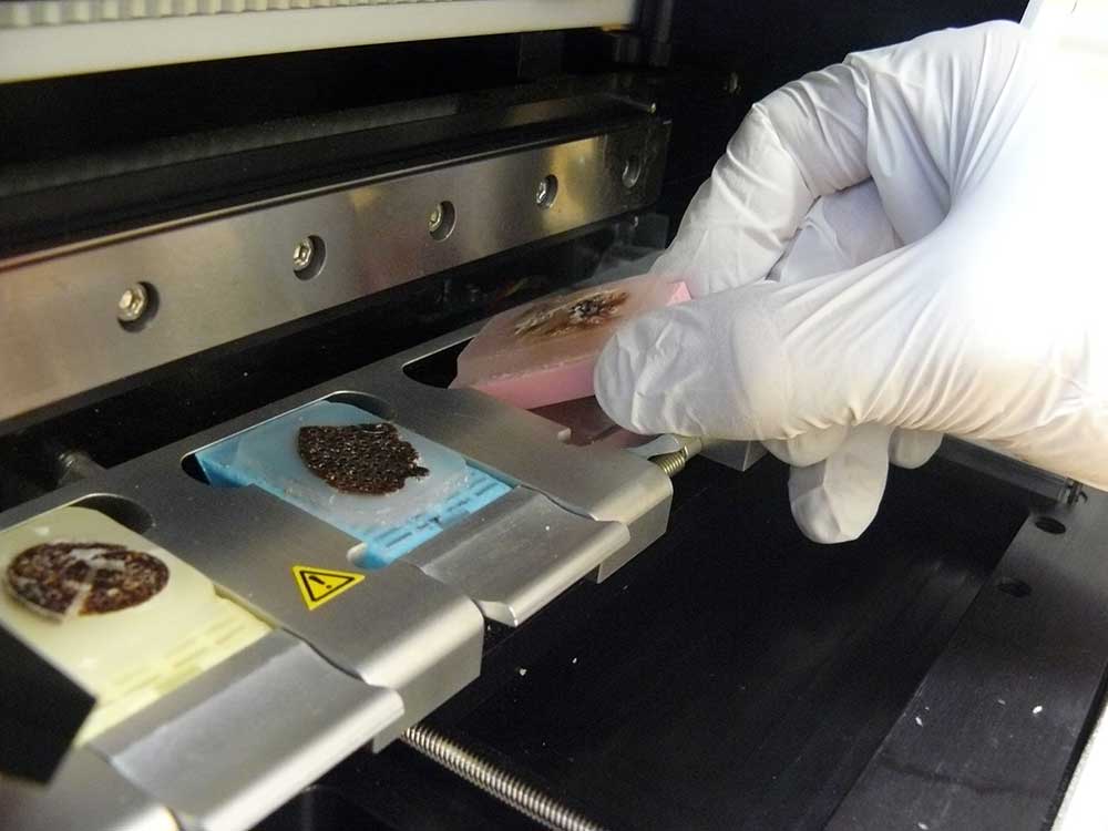
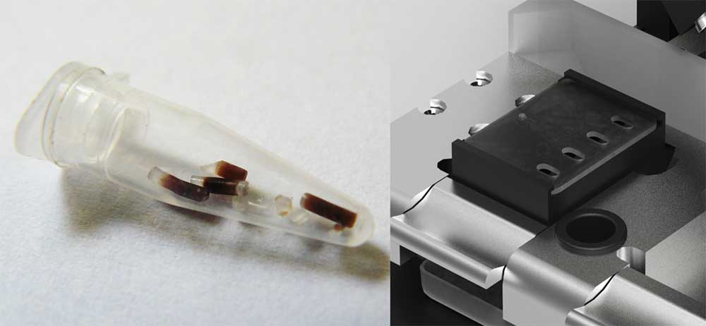
TMA Master II is a robust and reliable computer-controlled instrument for creating tissue microarray (TMA) blocks. It is an ideal solution for smaller laboratories, helping save reagent, slides, time, workforce and money. By using TMA Master II, you can easily and quickly create TMA blocks with great precision, of 0.6, 1, 1.5 and 2 mm diameter tissue cores. Optionally, the instrument can also extract tissue samples to standard 0.2 ml PCR tubes. The extracted tissue samples can then be used later for various applications at the field of molecular pathology.
TMA Master II prepares TMA blocks from biological specimens and samples of human or animal tissues. Typical applications include, but are not limited to the following:
- Laboratories at hospitals and clinics
- Pathology departments at hospitals and clinics
- Biobanks
- Research centers, in particular cancer research centers
- Pharmaceutical companies
- Toxico-pathology laboratories
This easy-to-use device is one of the most advanced tissue microarrayers on the market with an impressive feature set.
Key Features
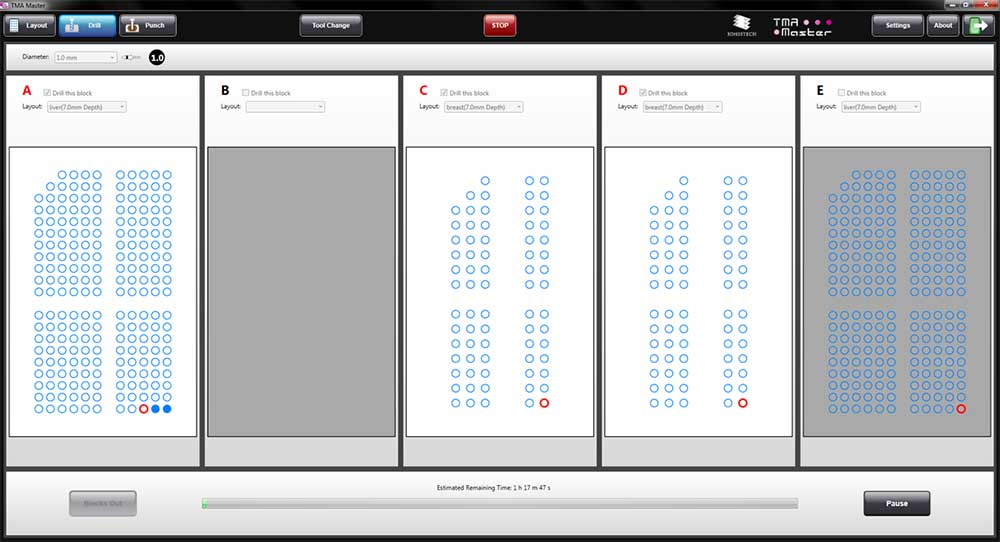
- Fully automated control
- Fast operation
- New, strong TMA Grand Master type motors and tools
- Automated tool size measurement for more precise operation
- Automated block height measurement for better TMA block quality
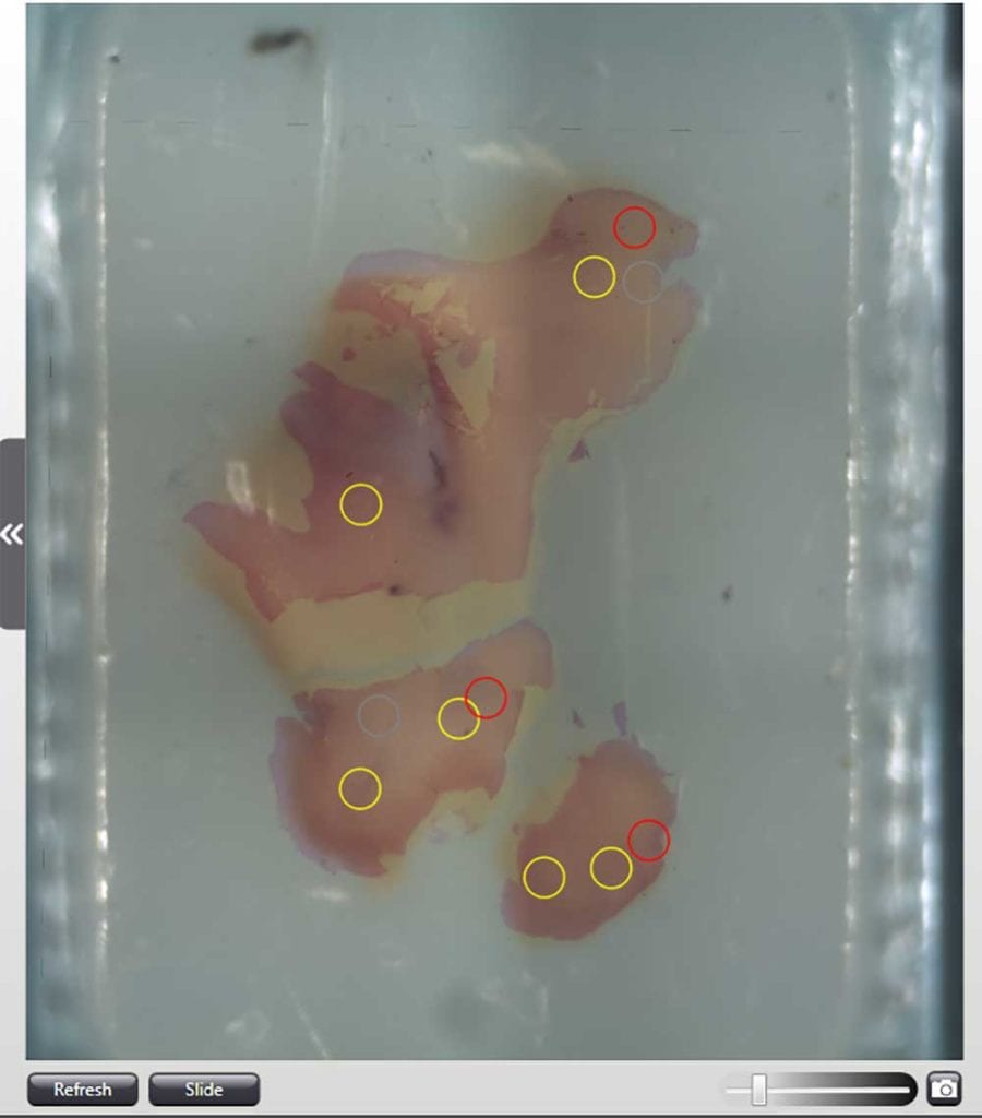
- New high resolution camera for donor block imaging
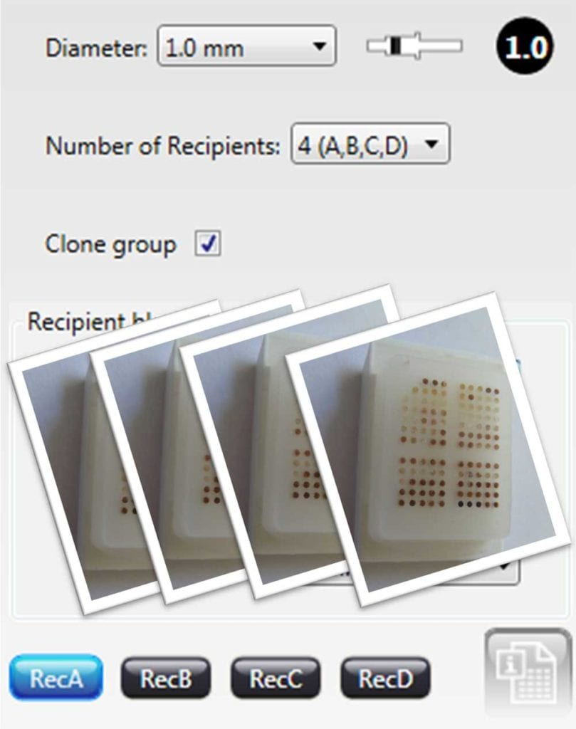
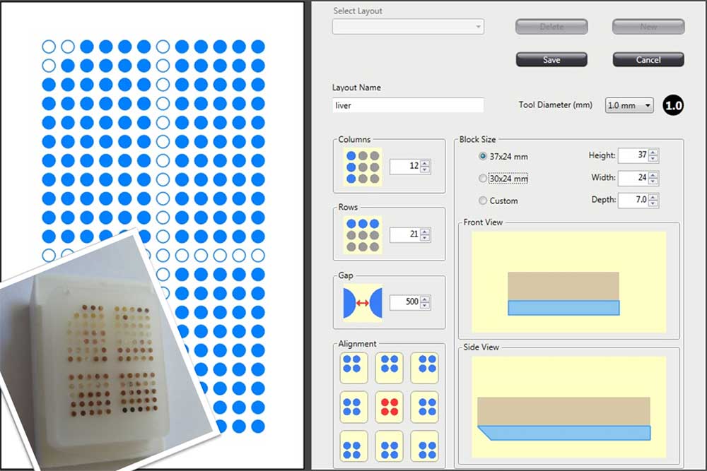
- Custom recipient block design and production
- Ability to create up to 4 identical TMA blocks
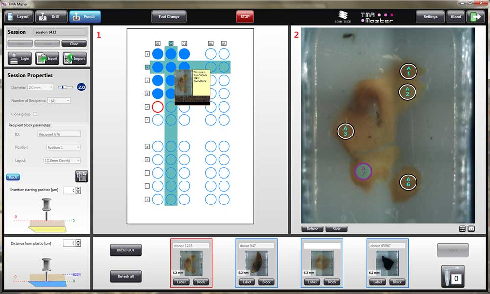
- Digital Slide overlay for more precise tissue core (sample) selection
- New PCR Cassette with 4 PCR tube capacity
- Sample extraction for molecular analysis
Software
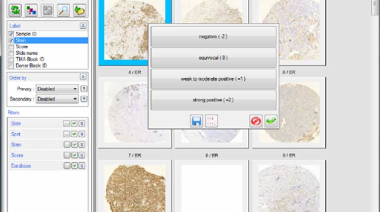
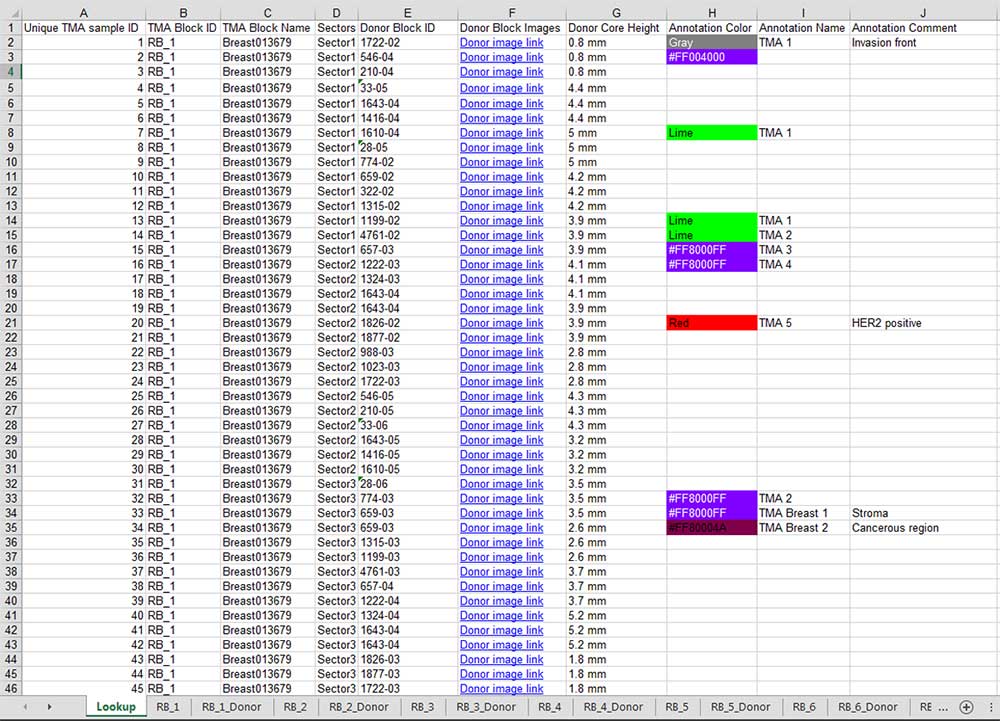
TMA Master II is controlled by the latest version of the TMA Control software. This software is the best solution for TMA block design and creation. It has advanced features for layout design and tissue core selection. A fully automated tool for overlapping an annotated digital slide with the donor block image helps precise core selection. During the TMA project the software saves automatically all the TMA data, which will be an invaluable source of information during TMA slide analysis using the TMA Module software.
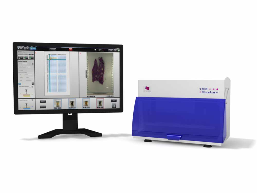
Specifications
| Capacity, blocks (Recipient and/or Donor) | 5 |
| Core diameter, mm | 0.6, 1, 1.5, 2 |
| Maximum number of cores |
558 (0.6 mm), 286 (1 mm), 135 (1.5 mm), 84 (2 mm) |
| Speed, core/hour | 200-250 |
| Donor block image recording | Yes |
| Use of MRXS digital slide and/or JPEG digital image for sample designation | Yes |
| Barcode reading | 1D and 2D |
| Data export | XLS |
| Dimensions, cm (W x D x H) | 38 x 24 x 29 |
| Weight, kgs | 8 |

