Pannoramic-X
Pannoramic-X: A New Era in 3D Digital Pathology
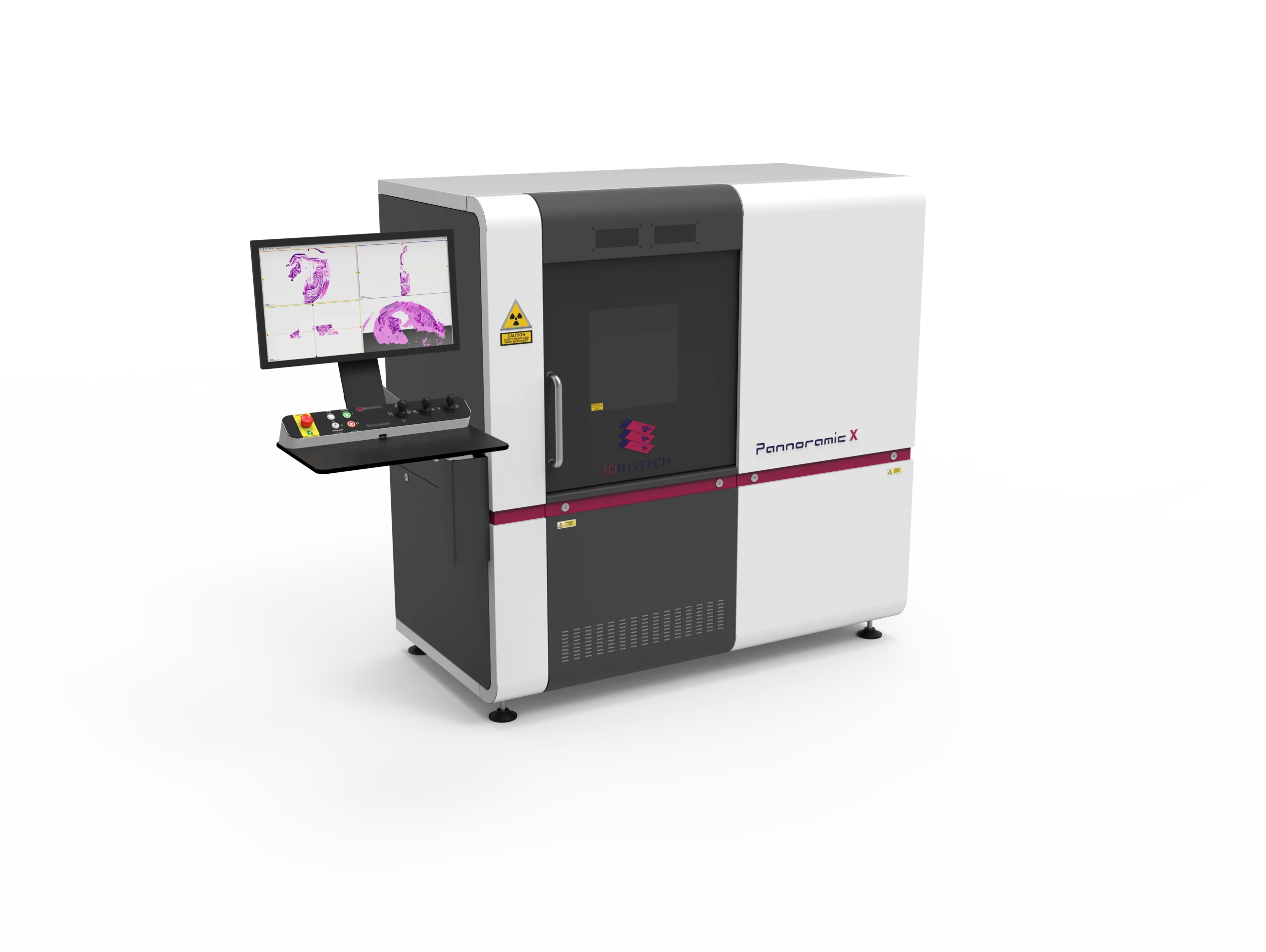
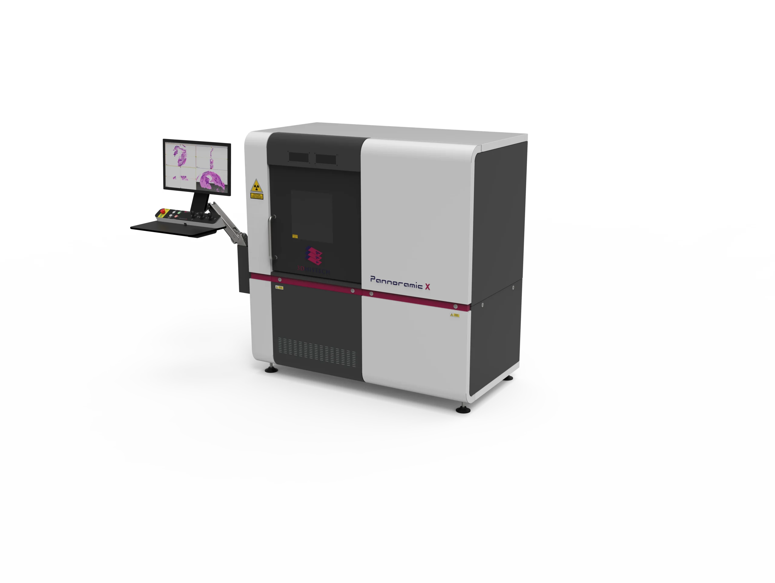
The Pannoramic-X micro-CT scanner revolutionizes 3D digital pathology by enabling ultra-high-resolution imaging of whole-FFPE tissue blocks, full blocks (macroblocks), and specimens up to 15 cm without traditional sectioning. Utilizing soft X-ray technology, it delivers virtual slicing and virtual staining, preserving sample integrity while enhancing biomarker analysis, tumor morphology assessment, and cancer staging. Powered by DVREAL 3D imaging, it provides unmatched depth and detail, surpassing conventional histology. Its intuitive software suite – CT Control, 3Dview, PatternQuant, and NuclearQuant – supports digital archiving, volumetric analysis, and AI-driven image processing for precise, reproducible results. With high-throughput capabilities, cost-effective operation, and integration into digital pathology workflows, the Pannoramic-X is an essential tool for cutting-edge research in cancer diagnostics, molecular pathology, and personalized medicine.
Unlike conventional slide-based histopathology, the Pannoramic-X eliminates depth and size limitations, accommodating large tissue volumes with high-detail scanning optimized for low-contrast samples. It provides an intuitive, user-friendly workflow, integrating seamlessly into digital pathology environments.
By combining unrivaled resolution, innovative digital histology, and AI-powered analysis, the Pannoramic-X is shaping the future of digital pathology, oncology research, and molecular diagnostics. It is a must-have for leading research institutions, pathology labs and pharmaceutical development centers aiming to push the bounderies of precision medicine and cancer diagnostics.
Key Features:
3D Digital Pathology Without Sectioning
Unlike traditional histology, which requires physical sectioning of FFPE blocks, the Pannoramic-X captures intact tissue structures in three dimensions using soft X-ray micro-CT technology. This eliminates the risk of tissue loss, distortion, or artifacts, preserving sample integrity for more accurate analysis.
Ultra-High-Resolution Imaging of Large Specimens
With the ability to scan whole-FFPE blocks, full blocks (Macro Blocks), and specimens up to 15 cm, the Pannoramic-X far surpasses traditional slide-based methods, which are limited by section size and depth. This expanded imaging capability ensures comprehensive tissue analysis for complex research applications.
Virtual Slicing & Staining with DVREAL Technology
The DVREAL 3D Imaging allows for virtual slicing and staining, replicating traditional histological techniques without physical alterations to the sample. This innovation enables multi-plane analysis, eliminates unnecessary cutting, and allows for retrospective examination of unstained tissue.
Enhanced Tumor Morphology & Biomarker Analysis
By providing detailed 3D visualization of tumor architecture, the Pannoramic-X enables precise assessment of lymphovascular invasion (LVI), tumor budding, and vascular structures – critical features for cancer staging and treatment planning. This level of detail is difficult to achieve with standard 2D slides.
AI-Powered Image Analysis for Faster, More Accurate Results
Integrated with PatternQuant and NuclearQuant, the Pannoramic-X supports high-speed, AI-driven image analysis, enabling automated pattern recognition, cell-based measurements, and quantitative biomarker assessments. This streamlines research workflows and ensures reproducible, unbiased data.
High-Volume, High-Throughput Scanning
Traditional pathology workflows involve manual sectioning, staining, and imaging, which can be labor-intensive and time-consuming. The Pannoramic-X offers high-throughput scanning, accommodating multiple samples simultaneously and reducing processing times.
Seamless Digital Pathology Integration
With CT Control and 3DView Software, the system integrates effortlessly into existing digital pathology infrastructures, supporting DICOM, MRXS, and RAW formats for compatibility with HIS and LIS systems. Researchers can store, analyze, and share 3D scans effortlessly.
Cost-Effective & Easy to Operate
Unlike traditional micro-CT systems that require specialized facilities, the Pannoramic-X is radiation-safe with zero emissions, operates on standard lab power, and requires no specialized engineers for maintenance. This reduces operational costs and expands accessibility.
| Feature | Pannoramic-X | Traditional Histology |
|---|---|---|
| 3D Imaging | Yes, with full volumetric analysis | No, limited to 2D slices |
| Sample Integrity | Intact, no sectioning required | Tissue loss and artifacts from cutting |
| Virtual Slicing & Staining | Yes, unlimited retrospective analysis | No, once sectioned, tissue is altered |
| Size Limitations | Scans up to 15 cm | Limited to thin sections |
| Tumor & Biomarker Analysis | Advanced 3D visualization and AI-driven quantification | Manual assessment from 2D slides |
| Workflow Efficiency | High-throughput, multiple samples per run | Labor-intensive, one slide at a time |
| Operational Cost | Cost-effective, no special facility needed | High cost due to labor and reagents |
- Micro-CT scanner
- Data Acquisition CPU (built-in),
- 4K Monitor, keyboard, and mouse (on the Arm)
- Reconstruction CPU, Keyboard, mouse
- 4K Monitor
Micro-focus 70kV, 7W (upgrade to 250W available in 2025)
- 3D reconstruction from 2D serial sections
- Advanced measurement tools for tumor morphology and biomarker quantification
- Color manipulation and adjustable viewing parameters
1790 mm x 929 mm x 1820 mm (without monitor console)
1150 kg (requires 2000 mm x 2800 mm floor space, below 500 kg/m²)
Enclosed, zero-emission system, requiring no special facility modifications
Operates on standard lab electricity, no specialized infrastructure needed
Scanning Capabilities
Sample Size
Few mm to 150 mm (15 cm)
X-ray Source
Micro-focus 70kV, 7W (upgrade to 250W available in 2025)
Scanning Time
Typically, 20 minutes to a few hours for high-resolution images
Operational Mode
24/7 continuous operation
Multiple Sample Loading
Supports simultaneous scanning of multiple samples (optional sample changer available)