06.03.2025 | Welcome to our brand new website!

TMA Master II

As the smallest fully automated tissue microarray system on the market, the TMA Master II optimizes laboratory space without compromising performance. Its compact footprint allows integration into any lab environment, enhancing workflow efficiency. Advanced automation ensures precise tool size and block height measurements, enabling reproducible array construction. With the capability to generate up to four identical TMA blocks and a dedicated PCR cassette for streamlined molecular analysis, it surpasses conventional systems by combining superior accuracy, reliability, and space efficiency for high-throughput research applications.
tma master ll
TMA Master II

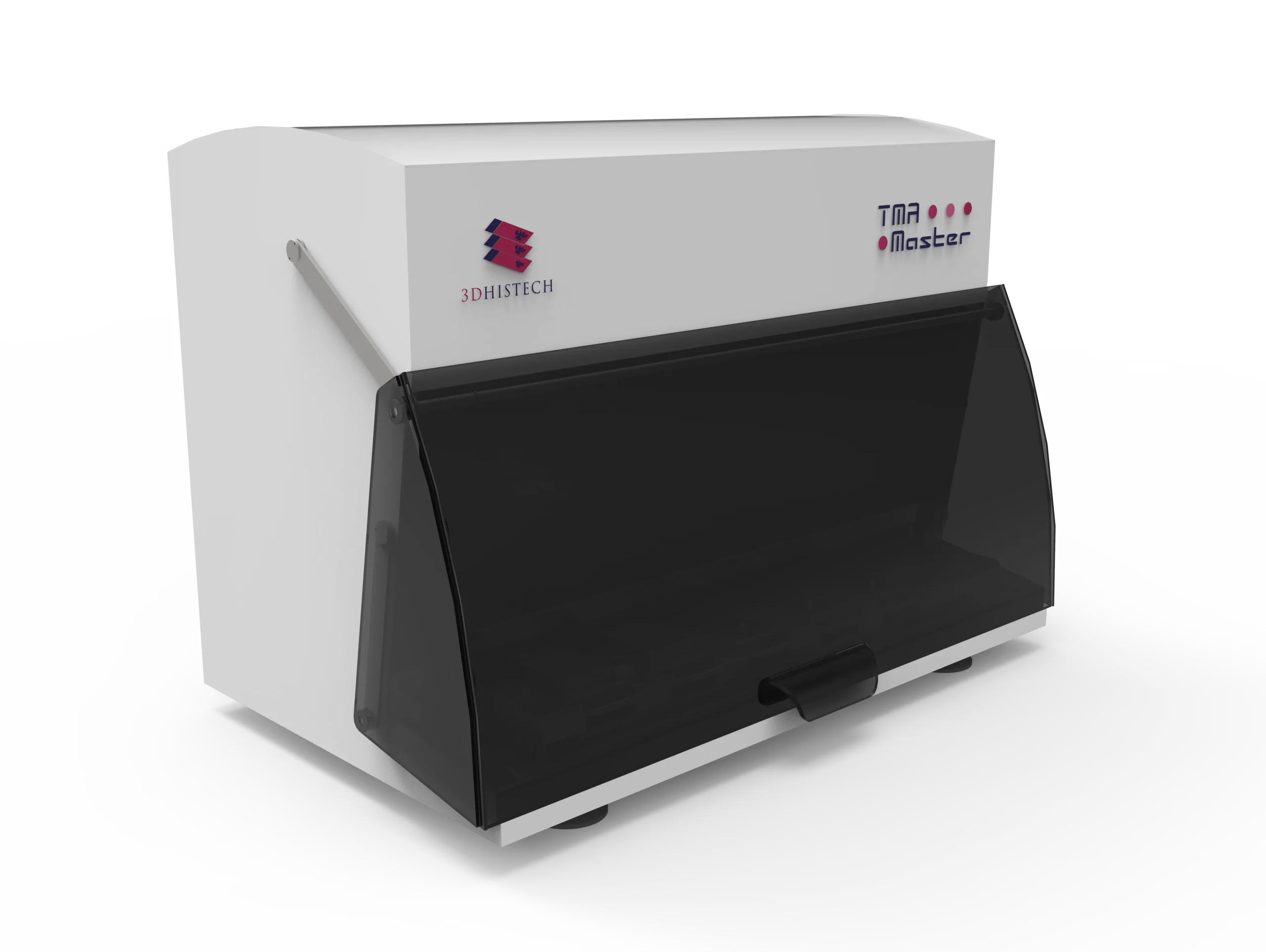
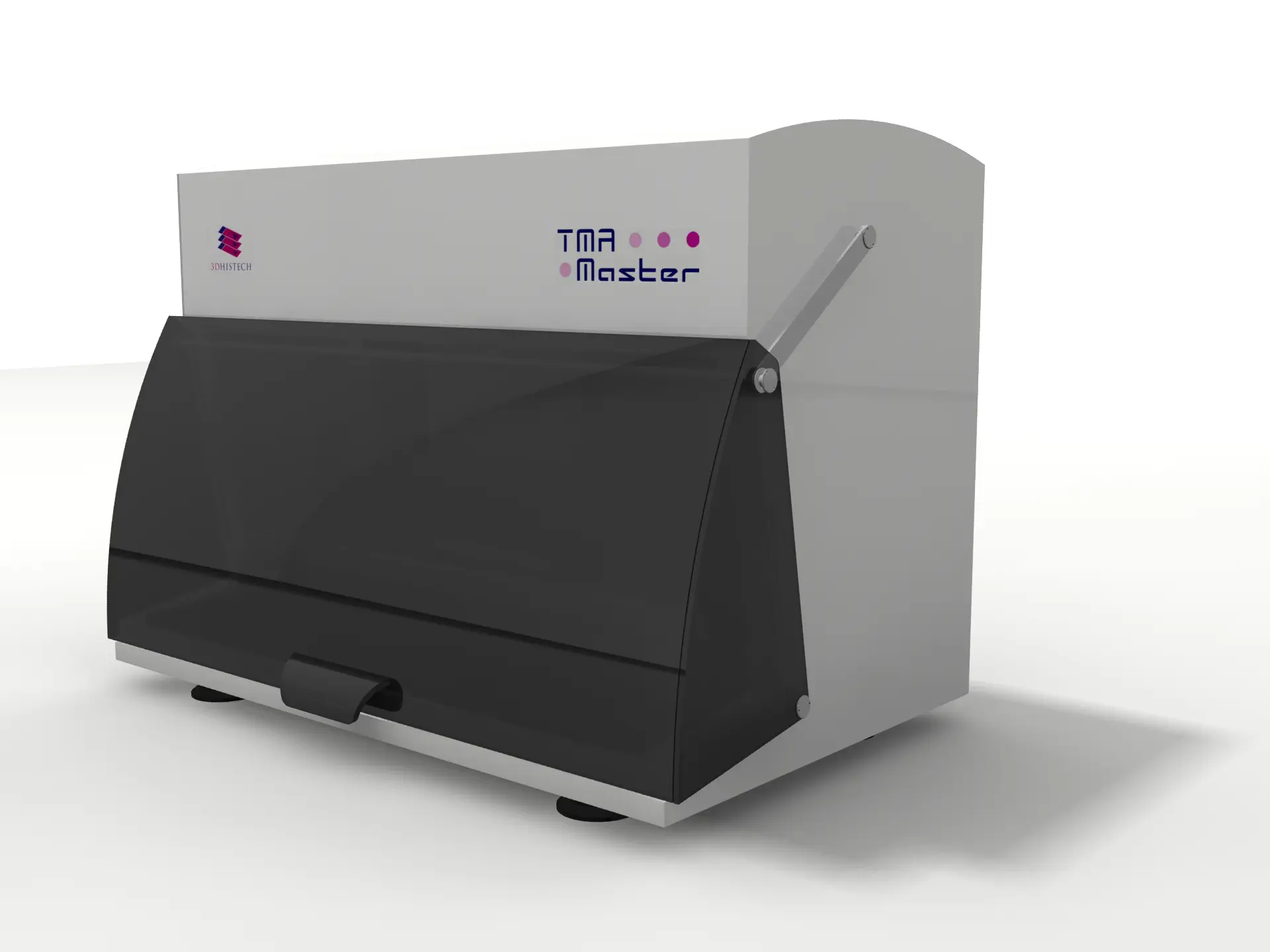
The TMA Master II – Precision Tissue Microarray in a Compact, Fully Automated solution

The TMA Master II is the most compact fully automated tissue microarrayer on the market, offering advanced functionality in a compact design that fits any laboratory bench. As an evolution of the TMA Master, it incorporates several hardware and software enhancements inspired by 3DHISTECH’s flagship TMA Grand Master, delivering improved precision, efficiency, and reliability.

Designed for pathology laboratories, biobanks, pharmaceutical research, and molecular diagnostics, the TMA Master II is a computer-controlled system that creates high-quality tissue microarray (TMA) blocks with 0.6, 1, 1.5, and 2 mm diameter cores. It can generate up to four identical TMA blocks, ensuring reproducibility for large-scale studies. Additionally, it includes an optional PCR cassette with four PCR tube capacity, allowing tissue sample extraction for molecular pathology applications.

Unlike conventional systems, the TMA Master II ensures superior precision and efficiency through features such as automated tool size and block height measurements, a high-resolution donor block camera, and digital slide overlay technology for precise tissue selection. With upgraded motors and tools from the TMA Grand Master, it operates at speeds of up to 250 cores per hour, making it one of the most efficient systems in its category.

Controlled by the latest TMA Control software, this system streamlines TMA block design, data management, and digital slide integration, ensuring accurate sample selection and documentation.

With its space-saving design, automation-driven precision, and optional molecular pathology capabilities, the TMA Master II is a powerful and cost-effective solution for laboratories seeking high-quality, reproducible TMA preparation in a compact system.

Key Features:

The Smallest Fully Automated Tissue Microarrayer
  • Requires minimal bench space, making it an ideal solution for laboratories with limited room.
  • Unlike larger systems, it provides high-throughput tissue microarray (TMA) block preparation without compromising laboratory space efficiency.
  • Reduces manual workload, minimizes human error, and ensures reproducible results.
  • Many competing systems require manual intervention, while the TMA Master II fully automates TMA block creation for consistent, high-quality output.
  • Saves time by processing high volumes of tissue cores efficiently.
  • Faster than many alternative systems, enhancing lab productivity and reducing turnaround time for research and diagnostics.
  • Provides greater durability, stability, and accuracy during sample extraction and core placement.
  • Utilizes proven high-performance motor technology from 3DHISTECH’s flagship TMA Grand Master for superior mechanical reliability.
  • Ensures consistent core dimensions and optimal block quality without manual calibration.
  • Eliminates variability, improving the reproducibility of TMAs for high-precision research.
  • Enhances imaging accuracy for sample selection and documentation.
  • Provides better visualization than standard systems, reducing the risk of errors in core placement.
  • Allows users to tailor block layouts for specific research needs.
  • Offers greater flexibility compared to fixed-template systems, accommodating diverse experimental designs.
  • Ensures sample consistency across multiple experiments, reducing the need for redundant tissue samples.
  • Many systems require separate processing for duplicate blocks, whereas TMA Master II generates identical TMAs simultaneously.
  • Integrates digital pathology tools to accurately align donor tissue sections with recipient blocks.
  • Reduces sample loss and improves targeting accuracy compared to manual selection methods.
  • Supports tissue sample extraction for molecular analysis, expanding research applications.
  • Many TMA systems lack integrated options for molecular pathology, making TMA Master II more versatile.
  • Automates TMA block design, sample tracking, and data management, saving time and improving reproducibility.
  • Features real-time data saving and automated digital slide integration, unlike many competing systems that rely on manual data entry.
Capacity & Throughput
  • Maximum Number of Blocks (Recipient and/or Donor): 5
  • Core Diameter Options: 0.6 mm, 1 mm, 1.5 mm, 2 mm
  • Maximum Number of Cores per Block:
  • 6 mm: 558 cores
  • mm: 286 cores
  • 5 mm: 135 cores
  • 0 mm: 84 cores
  • Processing Speed: 200-250 cores per hour
  • Ability to Create Identical TMA Blocks: Up to 4
Automation & Precision Features
  • Automated Tool Size Measurement: Yes
  • Automated Block Height Measurement: Yes
  • Custom Recipient Block Design & Production: Yes
  • Digital Slide Overlay for Tissue Core Selection: Yes
  • Donor Block Image Recording: High-resolution camera for precise imaging
Software & Data Management
  • Control Software: Latest version of TMA Control software
  • Data Export Format: XLS
  • Barcode Compatibility: 1D and 2D barcode reading
  • Sample Designation: Uses MRXS digital slide and/or JPEG digital images
Molecular Pathology Compatibility
  • PCR Cassette Capacity: 4 PCR tubes
  • Tissue Sample Extraction for Molecular Analysis: Optional
Physical Specifications
  • Dimensions (W x D x H): 38 x 24 x 29 cm
  • Weight: 8 kg
TMA Master II

Specifications

  • Instrument Type: Fully automated tissue microarrayer
  • Automation Level: Computer-controlled operation
  • Application: Tissue microarray (TMA) block creation from human or animal tissue samples
  • Laboratory Compatibility: Suitable for hospital pathology labs, research centers, biobanks, pharmaceutical companies, and toxico-pathology laboratories

Use Cases of TMA Master II

Explore the Use Cases

Archaeology and paleontology have traditionally relied on destructive sampling methods such as sectioning, staining, and thin-section petrography to study fossils and artifacts at the microscopic level. However, these approaches pose risks to…
Biomedical research is experiencing a major shift with the integration of Digital Pathology and Micro-CT solutions, significantly improving tissue analysis, biomarker discovery, and disease modeling. Traditional microscopy methods have inherent limitations, such…
Rapid and accurate biopsy diagnostics and frozen section evaluations are critical for clinical decision-making, especially in oncology and intraoperative pathology. 3DHISTECH’s Pannoramic DX digital pathology solutions provide a complete workflow for digital…
Botany and plant sciences rely on high-resolution imaging and precise analysis of plant tissues to understand disease mechanisms, stress responses, genetic modifications, and ecological changes. Traditional microscopy techniques limit research efficiency due…
Cancer research demands cutting-edge imaging technologies to enhance biomarker discovery, tumor profiling, and immuno-oncology advancements. Traditional histopathology techniques, while fundamental, present challenges such as limited scalability, subjective interpretation, and data fragmentation. Digital…
As pathology departments face increasing case volumes and demands for faster diagnoses, traditional workflows can struggle with time-intensive slide analysis, inconsistent imaging, and limited remote collaboration. Digital Pathology offers a modern solution,…
Cytopathology plays a critical role in diagnosing cancer, infectious diseases, and other cellular abnormalities. The transition from conventional slide review to digital pathology enhances efficiency, precision, and collaboration in cytological diagnostics….
Developmental biology is a rapidly evolving field that focuses on understanding how organisms grow, develop, and differentiate at the cellular and molecular levels. Researchers face key challenges such as capturing high-resolution images…
Immunohistochemistry (IHC) plays a critical role in cancer diagnostics, biomarker discovery, and personalized medicine, requiring high-precision imaging, consistent staining interpretation, and workflow efficiency. 3DHISTECH’s Pannoramic DX Whole Slide Imaging (WSI) solutions provide…
Toxicological pathology and neuropathology require high-precision, high-throughput imaging solutions to assess tissue morphology, detect subtle abnormalities, and streamline data-intensive workflows. 3DHISTECH’s Pannoramic DX Digital Scanners provide fully automated, highresolution whole slide imaging…
Accurate diagnosis and efficient workflow are critical in gastrointestinal (GI) and liver pathology, where identifying subtle tissue changes can determine patient outcomes. The Pannoramic DX Digital Pathology Solutions featuring the Pannoramic 250…
Genetic research demands precise imaging tools capable of capturing high-resolution cellular and molecular structures for in-depth analysis. 3DHISTECH’s Solutions for digital pathology supported by QuantCenter, QuantServer, SlideManager, and SlideCenter, provide an AI-driven,…
Geological and mineralogical research relies on high-resolution imaging techniques to analyze mineral composition, rock microstructures, and sedimentary formations. Traditional petrographic microscopy, while effective, is often limited by sample preparation constraints, depth resolution,…
Immunology and infectious disease research require precise cellular analysis, biomarker quantification, and advanced multiplex imaging to understand immune responses, pathogen-host interactions, and disease progression. Traditional histopathology methods are labor-intensive, slow, and prone…
Molecular cell biology research relies on high-resolution imaging and fluorescence capabilities to analyze cellular structures, molecular markers, and complex biological interactions. The integration of digital pathology solutions from 3DHISTECH—Pannoramic Confocal, Pannoramic 250…
Neuroscience research demands high-resolution imaging, precise molecular analysis, and largescale data management to study the complexity of the brain, nervous system, and neurological diseases. Traditional microscopy and pathology techniques often limit the…
The fight against cancer demands precision, efficiency, and collaboration in pathology. The Pannoramic DX Whole Slide Imaging Solutions, including the Pannoramic 250 Flash III DX, Pannoramic 150 DX, and Pannoramic 75 DX…
Pharmacology research is essential for drug discovery, preclinical evaluation, and biomarker validation. However, researchers face significant challenges in high-throughput tissue analysis, biomarker quantification, and data integration. Traditional microscopy and manual pathology workflows…
Advancing telepathology and remote consultation, the Pannoramic DX Digital Pathology Solutions provide an end-to-end digital pathology workflow, ensuring rapid, high-resolution scanning and seamless remote collaboration. These solutions support real-time diagnostics, expert consultations,…
Toxicology research is essential for understanding the effects of drugs, chemicals, and environmental pollutants on biological systems. Traditionally, toxicology studies rely on manual histological evaluation, which can be time-consuming, prone to subjectivity,…
Toxicology and environmental science research requires high-precision imaging, high-throughput analysis, and advanced molecular assessments to evaluate the impact of toxins, chemicals, and environmental pollutants on biological systems. Traditional microscopy and histopathology methods…
Veterinary pathology plays a crucial role in diagnosing diseases in animals, supporting clinical treatment, biomedical research, and toxicologic pathology. The Pannoramic DX Digital Pathology solutions from 3DHISTECH provide high-speed, high-resolution digital pathology…
Veterinary pathology plays a vital role in animal disease research, wildlife conservation, livestock farming, and food safety. As disease outbreaks, environmental changes, and food security challenges rise, digital pathology, AI-driven analysis, and…

Still thinking?

Ask us a question about TMA Master II

Ask us a question

Request Demo