Please, visit us at booth number 7 at the
Annual Meeting of the 25th European Congress of Pathology,
where 3DHISTECH proudly presents new developments.
Date: 31 August – 4 September, 2013
Location:Centro de Congressos de Lisboa, Lisbon, Portugal

by
Please, visit us at booth number 7 at the
Annual Meeting of the 25th European Congress of Pathology,
where 3DHISTECH proudly presents new developments.
Date: 31 August – 4 September, 2013
Location:Centro de Congressos de Lisboa, Lisbon, Portugal
by
Please, visit us at booth number 7 at the
Annual Meeting of the 25th European Congress of Pathology,
where 3DHISTECH proudly presents new developments.
Date: 31 August – 4 September, 2013
Location:Centro de Congressos de Lisboa, Lisbon, Portugal
by
You are invited to the next webinar of 3DHISTECH:
Please, register for one of the following sessions!
Date: 19th June, 2013.
Session 1,
9:00 CET
12:00 MSK
17:00 JST
19:00 AEDT
Register, Put it in my Calendar
Session 2,
8:00 PST
11:00 EST
14:00 BRT
17:00 CET
by
In the 1.15.4. release
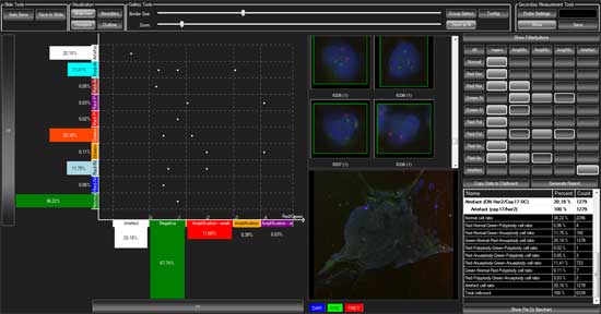
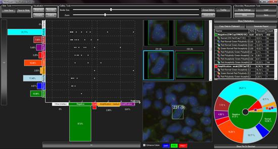
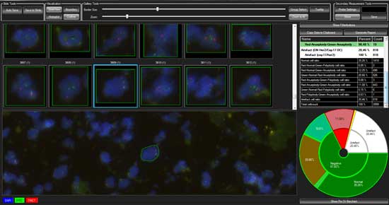
we made the measurement possibilities of FISHQuant complete. Not only
can FISHQuant work on cell samples, but the image processing of tissue
samples is also possible. The new program, while maintaining the
functionality of Cyto FISH, received a secondary non-specific
measurement option, which allows for the numerical analysis of
translocation probes, containing no structural abnormalities, and
display the combined results of the two measurements and statistical
analysis.
The new FISHQuant can be used for tissue and cytological
evaluation. Cytology samples which are cell cultures may contain cells
in metaphase whose identification and evaluation is done together with
the interphase cells as well as individually. Mitotic cells (metaphase)
in Cellcultures are also evaluated. The metaphase and interphase cells
results can be viewed together or separately.
• The algorithm can automatically identify the cells, and the signals,
• Probe specific Misp-file can be created for different samples containing the same marking,
• Users can select individual cells from a digital slide which can be evaluated in Quickmeasurement measurement mode
In many tumors, we encounter no specific deviations, but some numerical deviations . The program enables a secondary (numeric aberration) measurement method that identifies the numerical deviations of the candidate genes.
Histoplot: new visual display method for the amplification probes, in which the amplification rate group (gene / control mark =%) and the control gene marking numbers are displayed. It makes the reading of the aneoplid, polyploid populations amplification values easier.
Relationship between all populations, specific and non-specific (no difference) with one another to form easy to read display.
by
You are invited to the next webinar of 3DHISTECH:
“Reconstruct, Visualize and Measure
Histological Slides in 3 Dimensions with 3DView“,
Dr. Stephanie Starke presenting for MicroDimensions
Video: Kidney segmentation and glomerulus reconstruction in 3DView
Please, register for one of the following sessions!
Date: 28th May, 2013.
Session 1,
9:00 CET
12:00 MSK
17:00 JST
19:00 AEDT
Session 2,
8:00 PST
11:00 EST
14:00 BRT
17:00 CET
by
Please, be invited to visit booth number 416 at the Annual Meeting of the American Association for Cancer Research (AACR 2013), where 3DHISTECH proudly presents new developments.
Date: April 6-10th, 2013
Location: Walter E. Washington Convention Center, Washington, DC, USA
Exhibiting:
Laser microdissection is a widely utilized technology in life sciences research and for clinical applications. It is mainly used to accurately dissect diseased cells from healthy heterogeneous biopsy material. Laser micro-dissection is an essential tool for early diagnosis of cancers and neurological disorders.
According to the results of the 2nd International Scanner Contest (ISC), this is the fastest digital slide scanner on the market at both 20x and 40x resolution. Designed for routine digital pathology, the new Flash II easily meets your needs, from high quality to high capacity.
Experience the ultimate diagnostic environment for routine digital pathology, which brings you the comfort of the high resolution medical display and microscope-like navigation on digital slides.
Tissue microarray (TMA) machinery and software have been just renewed. PCR extraction is available both in the TMA Master and the TMA Grand Master, thus combines molecular analysis with histology diagnosis.
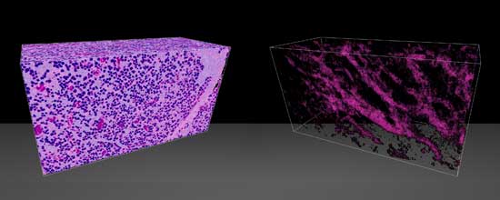
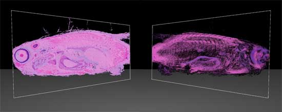
Microscope slides allow you to see one section of reality. Even with Z-stack or Extended focus, you are still constrained to that one section only. 3DView reconstructs the original specimen from its serial sections in minutes, thus lets you look into microscopic details while also showing you the tissue in its original form.
© 2005-2024, 3DHISTECH Ltd. All rights reserved.
H-1141 Budapest, Öv u. 3., Hungary
This website uses cookies so that we can provide you with the best user experience possible. Cookie information is stored in your browser and performs functions such as recognising you when you return to our website and helping our team to understand which sections of the website you find most interesting and useful.
Strictly Necessary Cookie should be enabled at all times so that we can save your preferences for cookie settings.
If you disable this cookie, we will not be able to save your preferences. This means that every time you visit this website you will need to enable or disable cookies again.
This website uses Google Analytics to collect anonymous information such as the number of visitors to the site, and the most popular pages.
Keeping this cookie enabled helps us to improve our website.
Please enable Strictly Necessary Cookies first so that we can save your preferences!