Key Challenges in Botany and Plant Sciences
Limited Resolution and Depth Perception in Plant Tissue Analysis
Traditional microscopy provides flat, 2D images that lack depth, limiting the study of internal cellular structures, vascular systems, and complex plant morphologies.
Time-Consuming & Labor-Intensive Slide Preparation
Preparing plant samples for microscopic analysis often requires thin sectioning, which is both time-consuming and destructive. Researchers also face challenges with fragile or delicate tissues.
Inefficiency in High-Volume Research
Large-scale studies, such as crop improvement and ecological research, require high-throughput scanning. Many existing solutions lack the capacity and automation needed for large sample processing.
Lack of Advanced Fluorescence Imaging for Plant Research
Photosynthesis studies, genetic engineering, and molecular plant research depend on fluorescence imaging to track protein expression and cellular interactions. Standard imaging systems fail to provide the required multi-channel fluorescence support.
Difficulty in Non-Destructive Plant Imaging
Some research applications require intact tissue analysis without slicing. Standard histological sectioning destroys samples, making retrospective analysis impossible.
optimizing botany and plant sciences
Whole Slide Imaging and 3D Digital Scanning for Plant Research
№1. Confocal Z-Stack scanning, 3D fluorescence imaging, minimal photobleaching
Resolves 2D imaging limitations, enhances fluorescence-based studies
Pannoramic Confocal Digital Scanner

№2. High-capacity (150 slides), brightfield and fluorescence imaging, 86x magnification
Ideal for high-throughput research and fluorescence-based applications
Pannoramic Scan II Digital Scanner

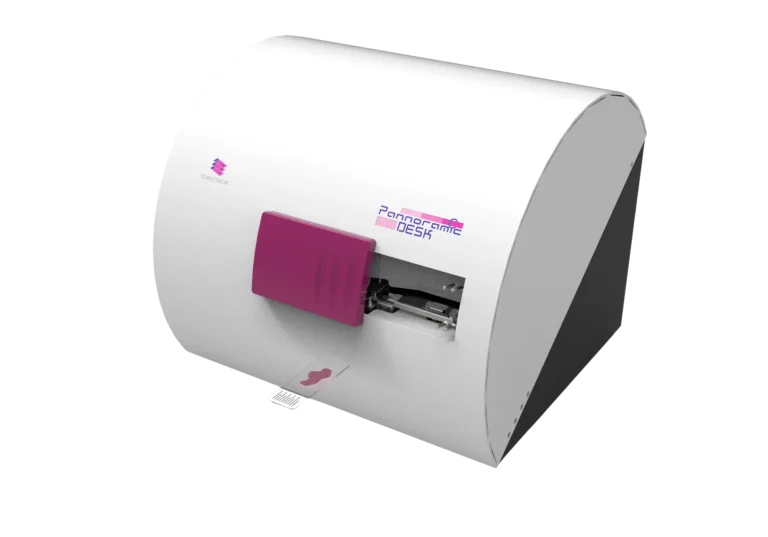
№3. Compact, high-resolution, brightfield imaging, Z-stack scanning
Suitable for small-scale labs, detailed analysis of complex plant tissues
Pannoramic Desk II DW Digital Scanner
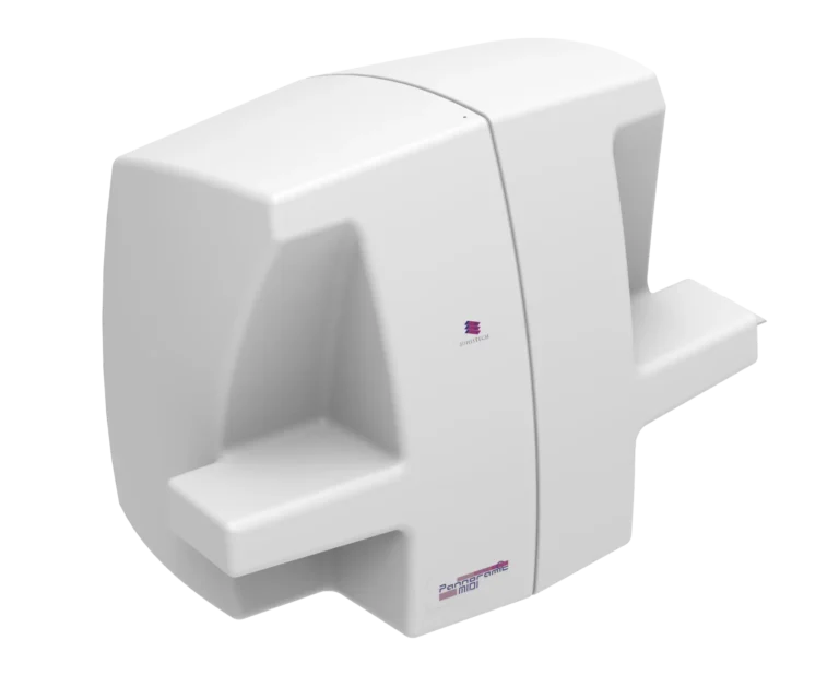
№4. Brightfield & fluorescence, 12-slide automation, Z-stack scanning
Supports fluorescence microscopy for plant biomarker studies
Pannoramic MIDI III Digital Scanner
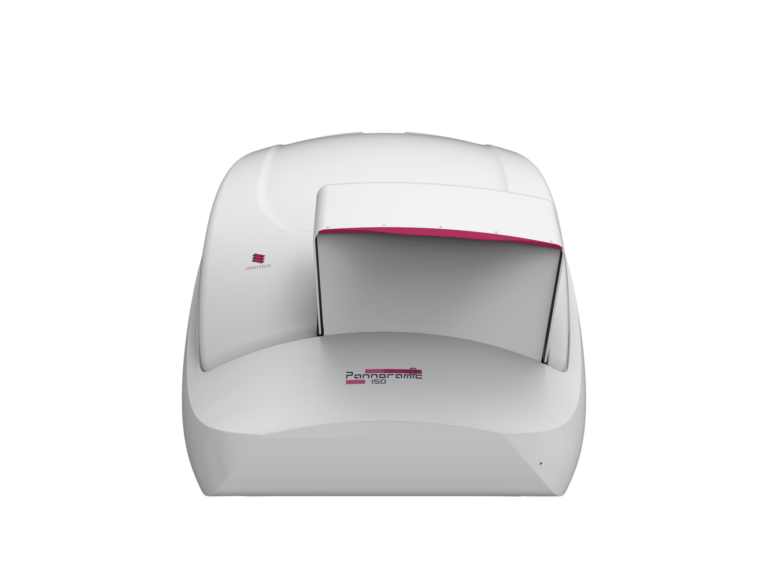
№5. 150-slide capacity, high-speed fluorescence scanning, AI-driven analysis
Best for high-throughput agricultural research
Pannoramic 150 Digital Scanner
№6. 3D digital imaging, virtual slicing, non-destructive analysis
Allows whole plant scanning without sectioning
Implementation
Use Case Scenarios
01. Disease Diagnosis in Agricultural Crops
Scenario
A plant pathology research lab investigates fungal infections in wheat crops to assess the impact of climate change on plant disease resistance. The research requires detailed imaging of fungal penetration in leaf tissues at a cellular level.
Implementation
- The Pannoramic Confocal Digital Scanner is used for fluorescence imaging, detecting fungal interactions with plant cells in high resolution.
- Researchers apply Z-stack scanning to create 3D reconstructions of infected tissues.
- Data is analyzed using QuantCenter (PatternQuant & HistoQuant) to quantify fungal spread and evaluate resistance levels in genetically modified crops.
Results & Impact
- The detection accuracy of fungal infections improved by 40% compared to traditional microscopy.
- Researchers identified key genetic markers that enhance plant immunity.
- The fluorescence imaging reduced sample processing time from 2 days to 3 hours.
02. High-Throughput Imaging for Crop Improvement Studies
Implementation
- The Pannoramic Confocal Digital Scanner is used for fluorescence imaging, detecting fungal interactions with plant cells in high resolution.
- Researchers apply Z-stack scanning to create 3D reconstructions of infected tissues.
- Data is analyzed using QuantCenter (PatternQuant & HistoQuant) to quantify fungal spread and evaluate resistance levels in genetically modified crops.
Results & Impact
- The detection accuracy of fungal infections improved by 40% compared to traditional microscopy.
- Researchers identified key genetic markers that enhance plant immunity.
- The fluorescence imaging reduced sample processing time from 2 days to 3 hours.
Scenario
A genetics research center studying genetic modifications in rice plants requires automated high-throughput imaging to compare morphological changes across thousands of plant tissue samples.
03. Non-Destructive Imaging for Ecological Research
Scenario
A plant pathology research lab investigates fungal infections in wheat crops to assess the impact of climate change on plant disease resistance. The research requires detailed imaging of fungal penetration in leaf tissues at a cellular level.
Implementation
- The Pannoramic 150 Digital Scanner (with 150-slide capacity) enables continuous scanning of genetically modified plant samples.
- SlideManager software organizes and tracks slide metadata for efficient data handling.
- Fluorescence multiplexing helps researchers study gene expression linked to drought resistance.
Results & Impact
- Image analysis using AI-powered QuantCenter helped detect subtle phenotypic variations between modified and wild-type plants.
- The findings contributed to the development of drought-resistant rice strains with higher yield potential.
- Scanning efficiency increased by 75%, enabling large-scale genetic studies.
Digital Solutions in Botany and Plant Sciences: A Critical Innovation for Modern Research
The integration of digital pathology solutions in botany and plant sciences has transformed plant research by enhancing image quality, fluorescence analysis, and high-throughput workflows.
Key Takeaways:
Pannoramic Confocal, Scan II, MIDI III, and 150 scanners provide fluorescence imaging for plant biomarker analysis.
Pannoramic-X Micro-CT scanner revolutionizes non-destructive 3D imaging, enabling whole-plant studies.
SlideManager and QuantCenter software ensure efficient data handling and AI-driven quantification.
Final Impact:
By adopting 3DHISTECH’s digital imaging solutions, botanists, plant pathologists, ecologists, and agricultural researchers achieve greater accuracy, efficiency, and scalability, paving the way for groundbreaking advancements in plant sciences.
Who Benefits from These Solutions?
Plant Pathologists
More precise disease identification, leading to better crop protection strategies.
Agricultural Researchers
High-throughput imaging accelerates genetic modification studies for improved crops.
Ecologists & Environmental Scientists
Non-destructive 3D imaging provides deeper insights into plant-environment interactions.
Biotechnologists
Fluorescence imaging enables detailed molecular analysis of plant genetics.
University & Research Institutions
Digital archives improve data reproducibility and collaboration between researchers globally.