This case study explores how these solutions enhance research workflows, addressing key challenges in molecular cell biology, and offering real-world applications that transform how scientists visualize and analyze cellular processes.
The Whole Slide Imaging Digital Pathology Solutions – Pannoramic Confocal, Pannoramic 250 Flash III, Pannoramic MIDI III, and Pannoramic 150 scanners, combined with research software, provide an advanced ecosystem for molecular cell biology. These solutions address key challenges in fluorescence imaging, high-throughput workflows, and digital pathology data management, revolutionizing molecular research across multiple disciplines.
Optimizing Molecular Cell Biology RESEARCH
Key Challenges in Molecular Cell Biology & How WSI Solutions Address Them
№1. Capturing High-Resolution, Multi-Layered Cellular Structures
The Pannoramic Confocal scanner provides structured illumination confocal imaging with Z-stack scanning (up to 100 optical sections) for 3D tissue reconstruction, ensuring clear visualization of cellular depth, eliminating out-of-focus blur, and improving contrast for biomarker studies.
Pannoramic Confocal Digital Scanner

Real-World Application:
In cancer research, 3D tumor profiling helps visualize invasion patterns and tumor microenvironments, enhancing precision in drug response studies.
№2. Handling High-Volume Research Workflows Efficiently
The Pannoramic 250 Flash III scanner enables high-throughput imaging with 300-slide capacity, AI-driven tissue detection, and continuous loading, allowing researchers to process extensive datasets efficiently.
Pannoramic 250 FLASH III Digital Scanner

Real-World Application:
A pharmacology lab conducting high-content drug screening can process hundreds of slides in a single workflow, reducing turnaround time for analyzing treatment responses.
№3. Fluorescence Imaging Complexity & Signal Sensitivity
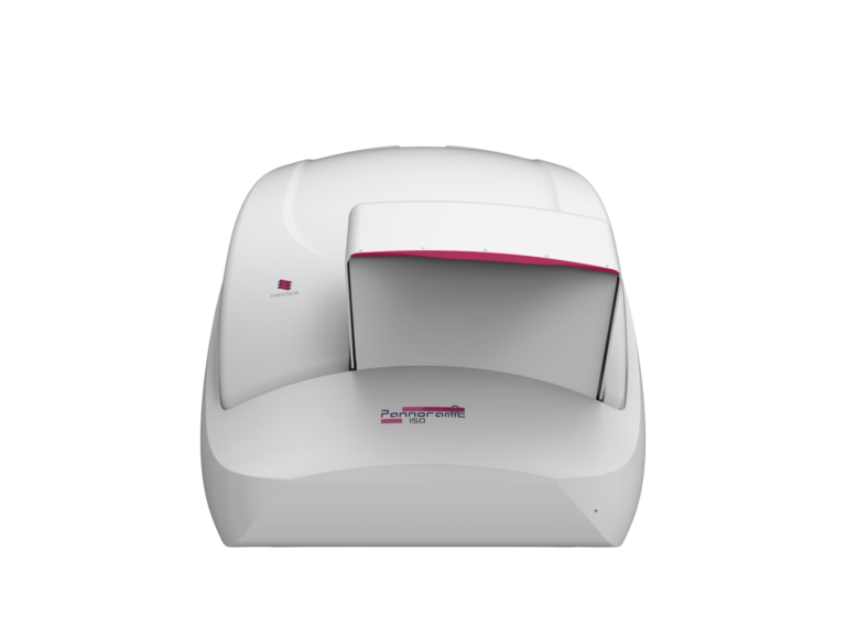
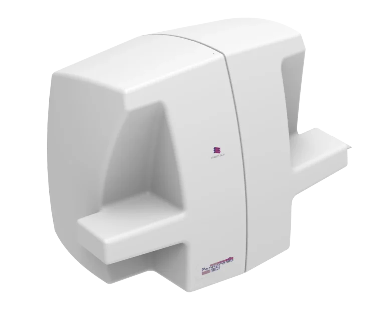
The Pannoramic MIDI III and Pannoramic 150 scanners feature high-end fluorescence imaging, with support for up to 45 fluorescent channels and automated Z-stack scanning for accurate biomarker visualization.
Pannoramic 150 Digital Scanner
Pannoramic MIDI III Digital Scanner
Real-World Application:
Neuroscientists studying synaptic plasticity use multiplex fluorescence imaging to quantify neural proteins across different brain regions, improving insights into neurodegenerative diseases.
№4. Data Management & Remote Collaboration
The SlideManager and SlideCenter platforms centralize research data, allowing multi-user access, remote collaboration, and cloud-based storage for secure data sharing across institutions.
№5. Quantitative Image Analysis for Molecular Research
QuantCenter and QuantServer enable AI-powered image analysis, including PatternQuant for pattern recognition and HistoQuant for automated biomarker quantification, ensuring reproducible results.
Real-World Application:
A multi-site cancer research consortium can share digital slide images for biomarker validation, facilitating seamless data exchange and interdisciplinary collaboration.
Who Benefits from Whole Slide Imaging Solutions in Molecular Biology?
Molecular Biologists & Pathologists
Utilize high-resolution imaging for molecular marker localization and cellular morphology analysis.
Cancer Researchers
Conduct biomarker quantification, study tumor microenvironments, and assess drug responses.
Neuroscientists
Use fluorescence-based imaging for brain tissue mapping and protein localization.
Pharmacologists
Perform high-throughput screening for drug discovery and toxicology assessments.
Immunologists
Analyze immune cell interactions and antigen expression.
Biotech & Pharma Labs
Require scalable digital solutions for research standardization and remote collaboration.
Transformative Applications of Whole Slide Imaging in Molecular Cell Biology
- Molecular Marker Localization Fluorescence scanning (45 channels) enhances biomarker visualization in cancer, neuroscience, and immunology research.
- Cell Morphology & 3D Tissue Reconstruction Confocal Z-stack scanning provides unparalleled cellular detail for pathology and regenerative medicine.
- High-Throughput Drug Screening Pannoramic 250 Flash III streamlines multi-slide scanning with AI-driven automation.
- Quantitative Image Analysis QuantCenter automates feature extraction, enhancing statistical accuracy in molecular studies.
- Remote Collaboration & Digital Research Archives SlideManager and SlideCenter allow research groups to share, annotate, and analyze digital slides worldwide.