Key Challenges in Gastrointestinal and Liver Diagnostics
Complex Tissue Structures
High cellular diversity and histopathological variations require precise imaging.
High Case Volumes
Large hospitals and diagnostic centers handle a significant number of GI and liver pathology cases, demanding high-throughput solutions.
Need for Remote Collaboration
Specialists often need second opinions and multidisciplinary consultations.
Regulatory and Compliance Requirements
Digital pathology systems must adhere to IVDR standards and integrate with LIS/HIS for streamlined case management.
Variability in Sample Types
Biopsies, resected tissues, and cytological specimens require adaptive scanning solutions.
Why Choose High-Throughput Scanners for GI and Liver Pathology?
Large Case Volume Handling
High-throughput scanning for rapid processing of large batches of histology and cytology slides.
Exceptional Image Quality
High-resolution scanning ensures accurate identification of subtle pathological changes in GI and liver tissues.
AI-Powered Automation
Reduces human intervention while improving diagnostic consistency.
Advanced Multi-Layer Imaging
Essential for cytology and challenging tissue structures.
Seamless LIS/HIS Integration
Ensures smooth workflow integration within hospital and laboratory systems.
Telepathology & Remote Consultation
Enables remote diagnosis and second opinions, vital for complex GI and liver pathology cases.
Optimizing Gastrointestinal and Liver Diagnostics
Solutions
By implementing the Pannoramic 1000 DX, Pannoramic 480 DX, and Pannoramic 250 Flash III
DX, medical institutions can modernize their pathology workflows, enhance diagnostic
precision, and ensure timely case processing. These scanners empower pathologists to deliver
high-quality patient care with confidence.
№1. Best for medium-to-large hospital and high-throughput GI pathology centers
- Optimized for High-Throughput, High-Resolution Imaging.
- AI-powered tissue detection for optimized scanning accuracy.
- Multilayer (Z-Stack) scanning for depth-enhanced imaging.
Key GI/Liver Use Cases:
- Histology-based diagnosis of liver cirrhosis, hepatitis, and GI malignancies.
- Cytopathology screening for fine-needle aspirates from liver or pancreas.
Pannoramic 250 Flash III DX Digital Scanner
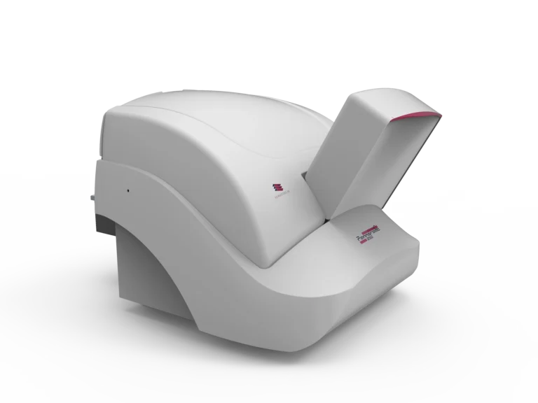
№2. Best for specialized GI and liver disease pathology departments, large-scale diagnostic hospitals
- High-Speed, Multi-Application Scanner with Polarization.
- Brightfield & Polarization Imaging – Essential for diagnosing birefringent materials like amyloid deposits in liver biopsies.
- Z-Stack and Extended Focus Imaging – Provides detailed depth analysis for challenging GI tissue structures.
- Safety Container – Separates problem slides without stopping the workflow.
- Touchscreen Interface for First-Level Quality Control.
Key GI/Liver Use Cases:
- Polarization imaging aids in diagnosing amyloidosis and liver fibrosis.
- 10x Objective High-speed scanning for large-scale liver and GI pathology screening
programs (scanning in half the time of traditional scanner units). - Integration with LIS and HIS.
Pannoramic 480 DX Digital Scanner
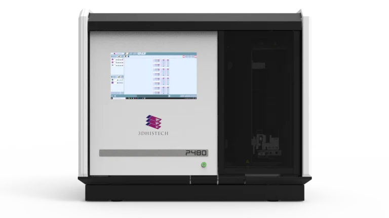
№3. Best for national-level pathology labs, high-volume liver disease research centers, and reference pathology institutions.
- The Ultimate High-Volume Scanner for GI and Liver Pathology
- High-Resolution Brightfield Imaging at 40x / 80x magnification.
- Z-Stack Imaging for Cytology, critical for detecting hepatocellular carcinoma and pancreatic cancer samples.
- AI-Driven Automated Workflow – Optimizes tissue recognition and slide quality control.
- Granite Base Anti-Vibration System – Ensures unparalleled imaging stability for highprecision diagnosis.
Key GI/Liver Use Cases:
- Advanced GI and liver histopathology for diagnostics.
- Ultra-high throughput for national cancer screening programs.
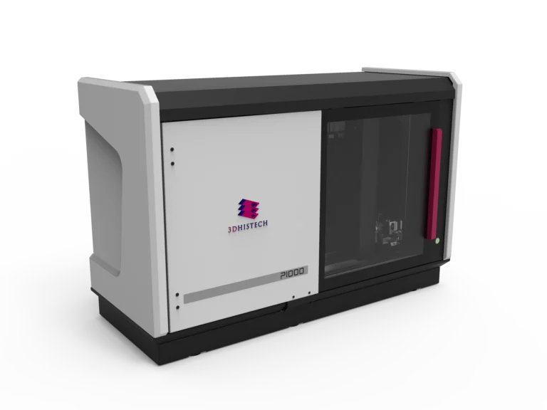
Pannoramic 1000 DX Digital Scanner
How the Pannoramic DX Solutions Address The Challenges
High-Resolution, AI-Powered Imaging
Pannoramic 250 Flash III DX:
- Ultra-fast scanning for large-volume facilities.
- AI-driven tissue detection for automated workflow.
- Multilayer scanning to capture fine tissue structures.
- Peripheral blood smear compatibility for additional hematological insights.
- Histology-based diagnosis of liver cirrhosis, hepatitis, and GI malignancies.
- Cytopathology screening for fine-needle aspirates from liver or pancreas
Pannoramic 150 DX:
- 0.1 um/pixel resolution at 100x magnification for highly detailed histological analysis.
- Fully automated slide handling with AI-assisted tissue and coverslip detection.
- DICOM and LIS/HIS integration for streamlined workflow.
Pannoramic 75 DX:
- Compact, high-throughput scanning for laboratories handling mid-range volumes.
- Z-stack and extended focus scanning for detailed morphological assessment.
- DICOM output compatibility, enabling seamless data exchange.
Enhanced Workflow and Remote Diagnostics
Telepathology Integration:
CaseManager DX allows specialists to collaborate remotely,
providing rapid second opinions on GI and liver cases.
Automated Workflow Optimization:
AI-powered quality control and barcode tracking
ensure seamless laboratory operations.
Fast Throughput:
Optimized for continuous slide scanning, reducing turnaround times.
Regulatory Compliance and Data Security
- IVDR-compliant software and hardware support clinical diagnostics in regulated environments.
- Secure Data Management: Integration with SlideStorage DX and SimpleSlideInterface ensures digital pathology images are safely stored and easily retrievable.
- DICOM-Ready Imaging: Ensures compatibility with existing PACS systems for effortless case management.
Optimizing Clinical Pathology
Why Choose 3DHISTECH’s Pannoramic DX Whole Slide Imaging Solutions?
Scalability
From small labs to large medical institutions, the Pannoramic DX scanners fit diverse workflow needs.
Uncompromised Imaging Accuracy
AI-powered tissue recognition and high-resolution scanning ensure diagnostic precision.
Seamless Integration
DICOM and LIS/HIS compatibility streamline digital pathology workflows.
Advanced Collaboration Tools
CaseManager DX enables remote diagnosis and consultations, improving patient outcomes.
Who Benefits from These Telepathology Solutions?
Gastroenterologists and Hepatologists
Gain accurate, high-resolution digital slides for diagnosing GI and liver diseases.
Pathologists
Leverage automated slide handling and AI-powered imaging to enhance efficiency.
Multidisciplinary Teams
Facilitate collaboration between radiologists, oncologists, and surgeons via telepathology solutions.
Hospitals and Diagnostic Laboratories
Improve diagnostic turnaround time with highthroughput, fully automated scanning.