06.03.2025 | Welcome to our brand new website!

TMA Grand Master

The TMA Grand Master automates tissue microarray creation with exceptional speed, precision, and reliability. Designed for high-throughput institutions, it ensures accurate core selection and placement, reducing manual effort while increasing reproducibility. With advanced features and intuitive operation, it outperforms traditional methods and competing systems in efficiency and consistency, making it the ultimate tool for institutional pathology labs, biobanks, large research centers, and pharmaceutical companies.
tma grand master
TMA Grand Master

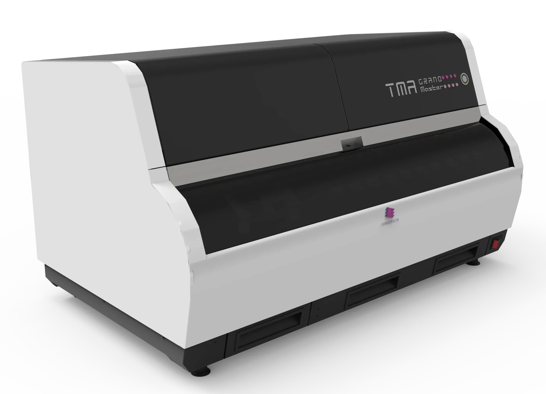
TMA Grand Master – The Ultimate High-Throughput TMA

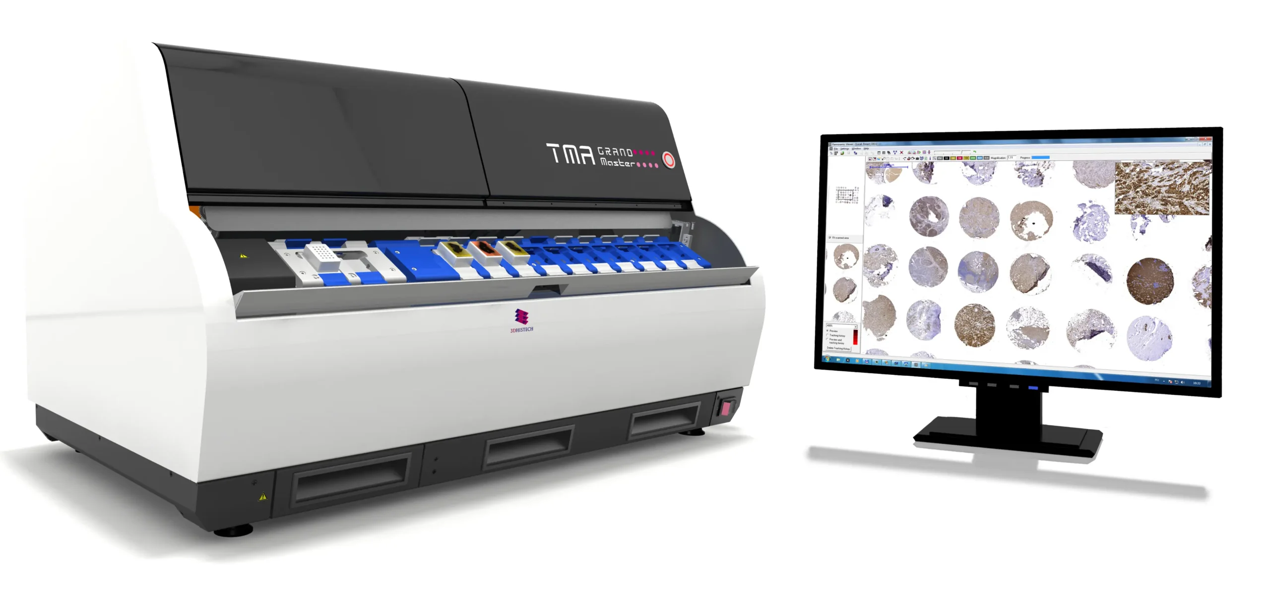
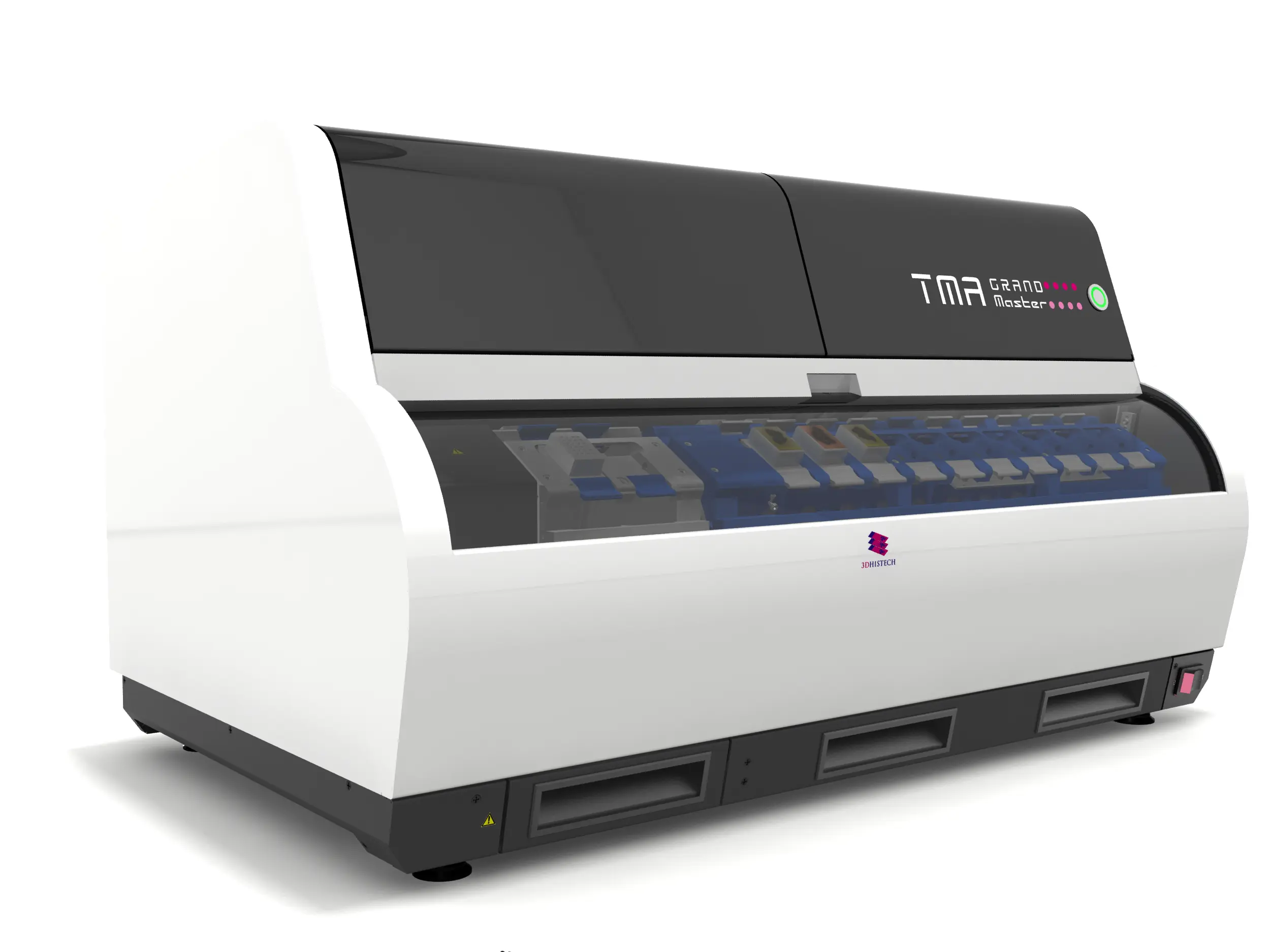
The TMA Grand Master is the industry-leading automated tissue microarrayer in the market, designed to streamline high-throughput tissue microarray (TMA) creation with unmatched precision, speed, and reliability. Engineered for research, biomarker studies, and drug development, it automates the selection, coring, and placement of tissue samples, significantly reducing manual workload while ensuring exceptional reproducibility. With its high-capacity design, the TMA Grand Master can process up to 72 recipient blocks and 600 donor blocks in a single run, far surpassing traditional methods and competing systems.

Its intelligent software allows for precise core selection, automated tissue recognition, and real-time visualization, minimizing human error and maximizing efficiency. The system’s high-speed robotic arm ensures accurate and consistent placement, while its barcode-based tracking enhances sample traceability. Designed for laboratories that demand both scalability and reliability, the TMA Grand Master sets the standard in automated TMA production – Featuring fully automated walkaway operation, 72 blocks capacity, 4 core diameters, ability to create clone blocks (identical TMAs), sample extraction feature for molecular analysis in PCR tubes (6 PCR casette, 10 PCR tube in each).

Key Features:

Market-Leading Speed and Capacity
  • Fastest fully automated tissue microarrayer available
  • Processes 72 blocks simultaneously (60 donor, 12 recipient)
  • High-speed operation – Processes up to 280 cores per hour
  • Four core diameters: 0.6mm, 1mm, 1.5mm, and 2 mm
  • High-density arraying: Up to 558 cores per block (0.6mm cores)
  • Custom recipient block design for tailored applications
  • Digital slide overlay for precise tissue core selection
  • Automatic block height measurement for perfect alignment
  • Integrated barcode scanning (1D and 2D) for efficient tracking
  • Automatic donor block and label image saving for documentation
  • Project data automatically saved to database for seamless analysis
  • PCR extraction function for downstream molecular pathology applications
  • Accommodates 6 PCR cassettes (each holding 10 PCR tubes)
  • FFPE tissue samples ready for DNA extraction and PCR analysis
  • Cleaning block integration to prevent cross-contamination
  • Imports clinical data and integrates with research workflows
  • Exports TMA data in multiple formats (ODS, XLS, XLSX, CSV, XML)
  • Compatible with digital pathology workflows, including MRXS and JPEG image formats
  • TMA Control software enables intuitive block design and tissue selection
  • Automated digital slide alignment ensures precise core placement
  • Comprehensive project tracking and TMA data management for streamlined analysis


Software & Workflow Integration
  • Total block capacity: 72 blocks (60 donor, 12 recipient)
  • Maximum core throughput: 250-280 cores per hour
  • Maximum cores per TMA block:
  • 6 mm cores: 558
  • mm cores: 286
  • 5 mm cores: 135
  • 0 mm cores: 84
Precision & Core Selection
  • Core diameter options: 0.6 mm, 1 mm, 1.5 mm, 2 mm
  • Automated donor block image recording for precise tissue selection
  • Digital slide integration: Supports MRXS and JPEG formats for sample designation
  • Fully automated barcode scanning: 1D and 2D
Automation & Data Management
  • Parallel processing: Simultaneous block loading, imaging, drilling, and punching
  • Automated block height measurement ensures perfect core alignment
  • Project data automatically saved to the internal database
  • TMA data export formats: ODS, XLS, XLSX, CSV, XML
Molecular Pathology Support
  • PCR extraction function:
  • 6 PCR cassettes, each holding 10 PCR tubes
  • Extracted FFPE tissue samples ready for DNA extraction and PCR analysis
  • Cleaning block integration to prevent cross-contamination
Physical Specifications
  • Dimensions (W x D x H): 80 x 50 x 46 cm
  • Weight: 48 kg


Software & Workflow Integration
  • Control Software: TMA Grand Master is powered by the latest TMA Control software, designed for precise block layout, core selection, and digital slide overlay.
  • Seamless workflow management: Automatically saves all TMA data for streamlined analysis with TMA Module software.
TMA Grand Master

Specifications

Use Cases of TMA Grand Master

Explore the Use Cases

Archaeology and paleontology have traditionally relied on destructive sampling methods such as sectioning, staining, and thin-section petrography to study fossils and artifacts at the microscopic level. However, these approaches pose risks to…
Biomedical research is experiencing a major shift with the integration of Digital Pathology and Micro-CT solutions, significantly improving tissue analysis, biomarker discovery, and disease modeling. Traditional microscopy methods have inherent limitations, such…
Rapid and accurate biopsy diagnostics and frozen section evaluations are critical for clinical decision-making, especially in oncology and intraoperative pathology. 3DHISTECH’s Pannoramic DX digital pathology solutions provide a complete workflow for digital…
Botany and plant sciences rely on high-resolution imaging and precise analysis of plant tissues to understand disease mechanisms, stress responses, genetic modifications, and ecological changes. Traditional microscopy techniques limit research efficiency due…
Cancer research demands cutting-edge imaging technologies to enhance biomarker discovery, tumor profiling, and immuno-oncology advancements. Traditional histopathology techniques, while fundamental, present challenges such as limited scalability, subjective interpretation, and data fragmentation. Digital…
As pathology departments face increasing case volumes and demands for faster diagnoses, traditional workflows can struggle with time-intensive slide analysis, inconsistent imaging, and limited remote collaboration. Digital Pathology offers a modern solution,…
Cytopathology plays a critical role in diagnosing cancer, infectious diseases, and other cellular abnormalities. The transition from conventional slide review to digital pathology enhances efficiency, precision, and collaboration in cytological diagnostics….
Developmental biology is a rapidly evolving field that focuses on understanding how organisms grow, develop, and differentiate at the cellular and molecular levels. Researchers face key challenges such as capturing high-resolution images…
Immunohistochemistry (IHC) plays a critical role in cancer diagnostics, biomarker discovery, and personalized medicine, requiring high-precision imaging, consistent staining interpretation, and workflow efficiency. 3DHISTECH’s Pannoramic DX Whole Slide Imaging (WSI) solutions provide…
Toxicological pathology and neuropathology require high-precision, high-throughput imaging solutions to assess tissue morphology, detect subtle abnormalities, and streamline data-intensive workflows. 3DHISTECH’s Pannoramic DX Digital Scanners provide fully automated, highresolution whole slide imaging…
Accurate diagnosis and efficient workflow are critical in gastrointestinal (GI) and liver pathology, where identifying subtle tissue changes can determine patient outcomes. The Pannoramic DX Digital Pathology Solutions featuring the Pannoramic 250…
Genetic research demands precise imaging tools capable of capturing high-resolution cellular and molecular structures for in-depth analysis. 3DHISTECH’s Solutions for digital pathology supported by QuantCenter, QuantServer, SlideManager, and SlideCenter, provide an AI-driven,…
Geological and mineralogical research relies on high-resolution imaging techniques to analyze mineral composition, rock microstructures, and sedimentary formations. Traditional petrographic microscopy, while effective, is often limited by sample preparation constraints, depth resolution,…
Immunology and infectious disease research require precise cellular analysis, biomarker quantification, and advanced multiplex imaging to understand immune responses, pathogen-host interactions, and disease progression. Traditional histopathology methods are labor-intensive, slow, and prone…
Molecular cell biology research relies on high-resolution imaging and fluorescence capabilities to analyze cellular structures, molecular markers, and complex biological interactions. The integration of digital pathology solutions from 3DHISTECH—Pannoramic Confocal, Pannoramic 250…
Neuroscience research demands high-resolution imaging, precise molecular analysis, and largescale data management to study the complexity of the brain, nervous system, and neurological diseases. Traditional microscopy and pathology techniques often limit the…
The fight against cancer demands precision, efficiency, and collaboration in pathology. The Pannoramic DX Whole Slide Imaging Solutions, including the Pannoramic 250 Flash III DX, Pannoramic 150 DX, and Pannoramic 75 DX…
Pharmacology research is essential for drug discovery, preclinical evaluation, and biomarker validation. However, researchers face significant challenges in high-throughput tissue analysis, biomarker quantification, and data integration. Traditional microscopy and manual pathology workflows…
Advancing telepathology and remote consultation, the Pannoramic DX Digital Pathology Solutions provide an end-to-end digital pathology workflow, ensuring rapid, high-resolution scanning and seamless remote collaboration. These solutions support real-time diagnostics, expert consultations,…
Toxicology research is essential for understanding the effects of drugs, chemicals, and environmental pollutants on biological systems. Traditionally, toxicology studies rely on manual histological evaluation, which can be time-consuming, prone to subjectivity,…
Toxicology and environmental science research requires high-precision imaging, high-throughput analysis, and advanced molecular assessments to evaluate the impact of toxins, chemicals, and environmental pollutants on biological systems. Traditional microscopy and histopathology methods…
Veterinary pathology plays a crucial role in diagnosing diseases in animals, supporting clinical treatment, biomedical research, and toxicologic pathology. The Pannoramic DX Digital Pathology solutions from 3DHISTECH provide high-speed, high-resolution digital pathology…
Veterinary pathology plays a vital role in animal disease research, wildlife conservation, livestock farming, and food safety. As disease outbreaks, environmental changes, and food security challenges rise, digital pathology, AI-driven analysis, and…

Still thinking?

Ask us a question about TMA Grand Master

Ask us a question

Request Demo