06.03.2025 | Welcome to our brand new website!

TMA molmed

Discover the TMA molmed, a fully automated, high-speed solution for tissue microarray creation and molecular sampling from FFPE and mega blocks. Combing precision microarraying with laser microdissection-like functionality, it enables highly accurate tissue extraction for advanced molecular applications, making it an essential tool for pathology, biobanking, and traslational research.
tma molmed
TMA molmed

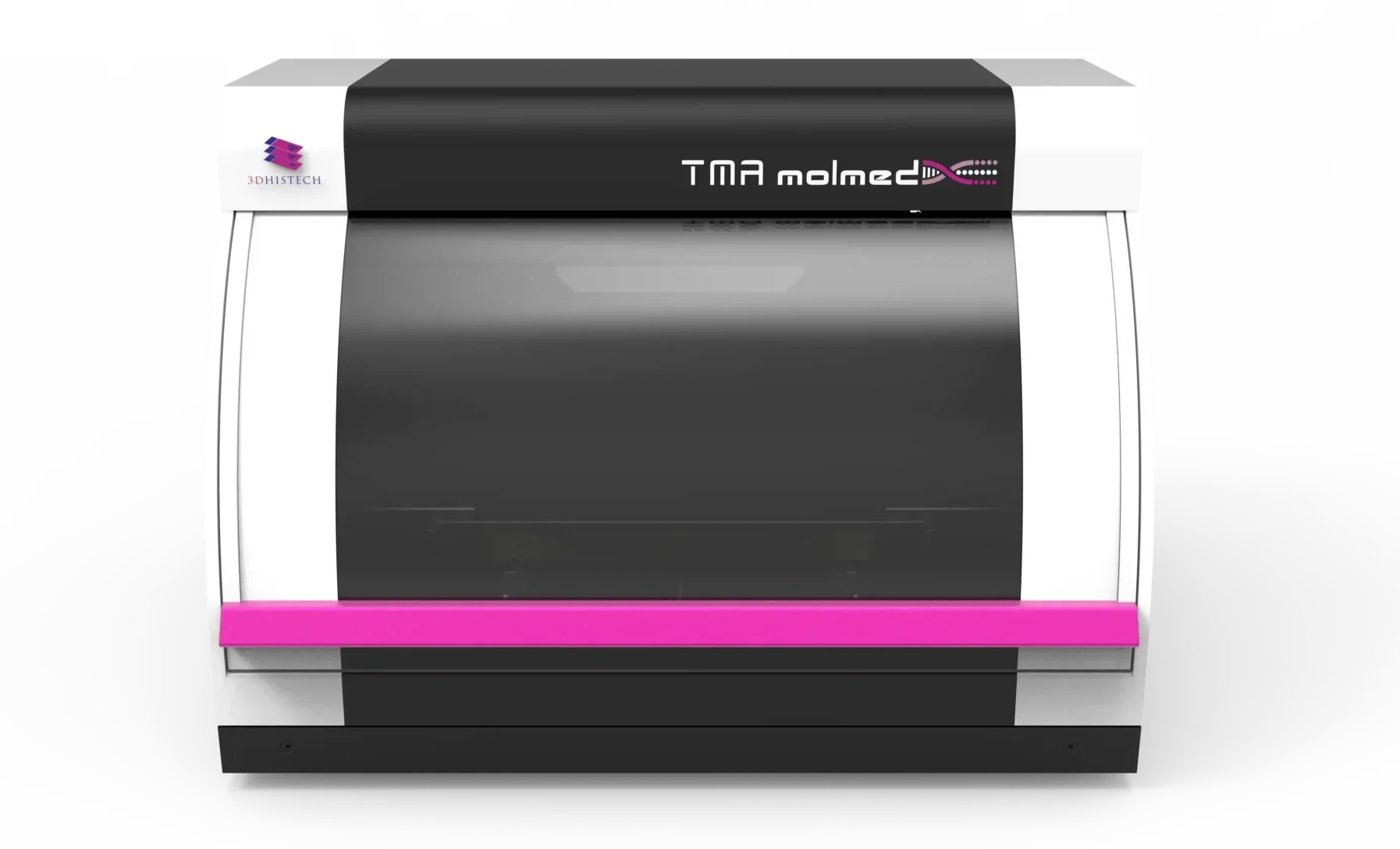
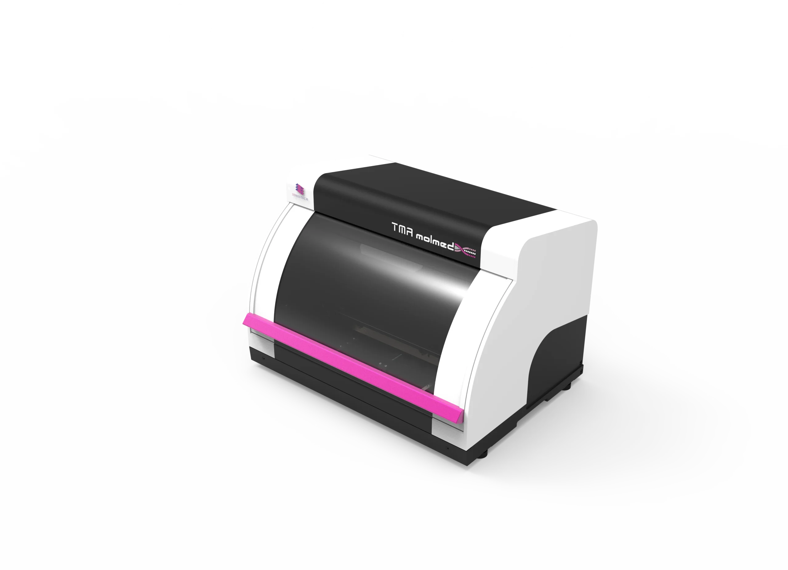
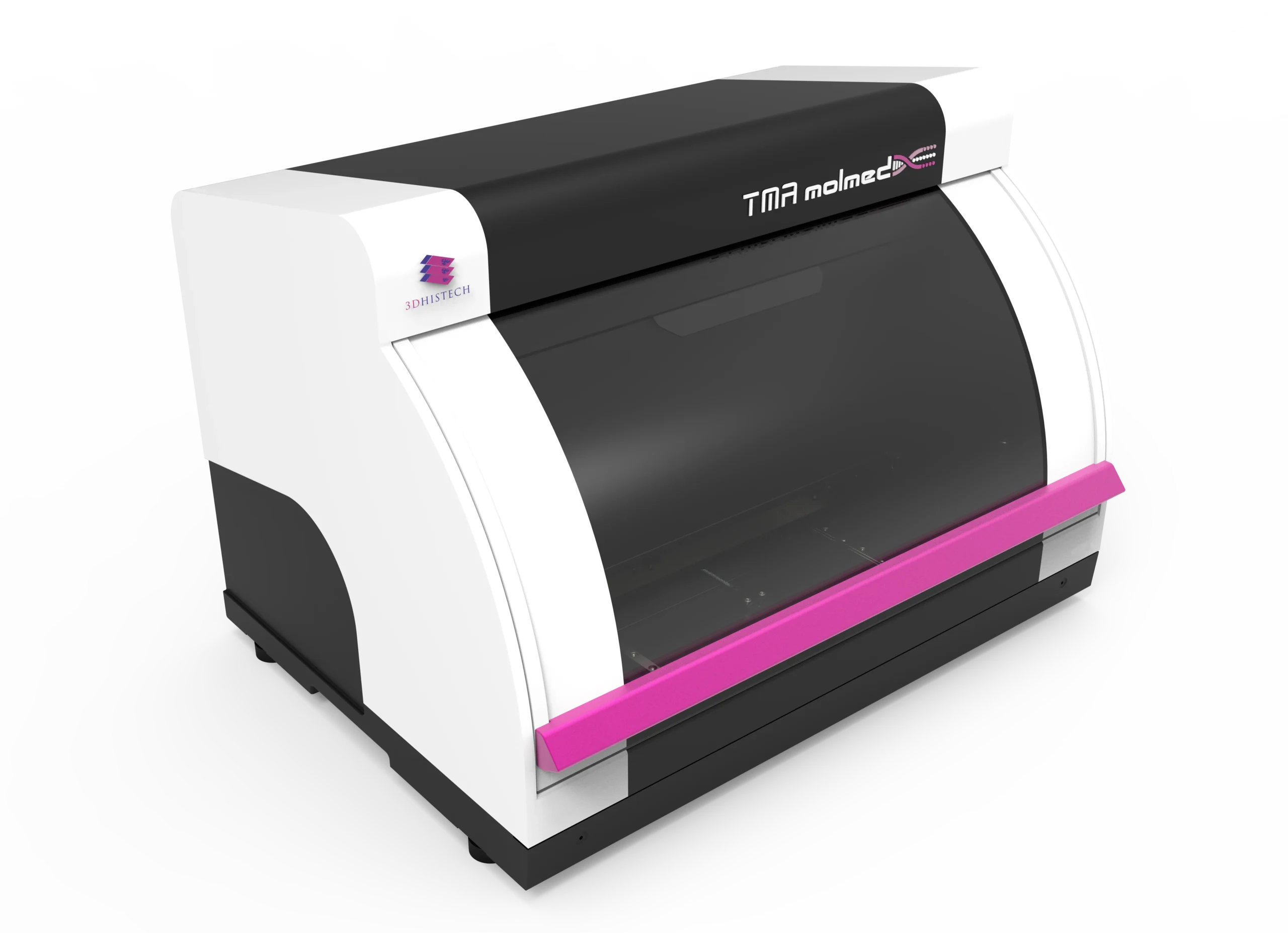
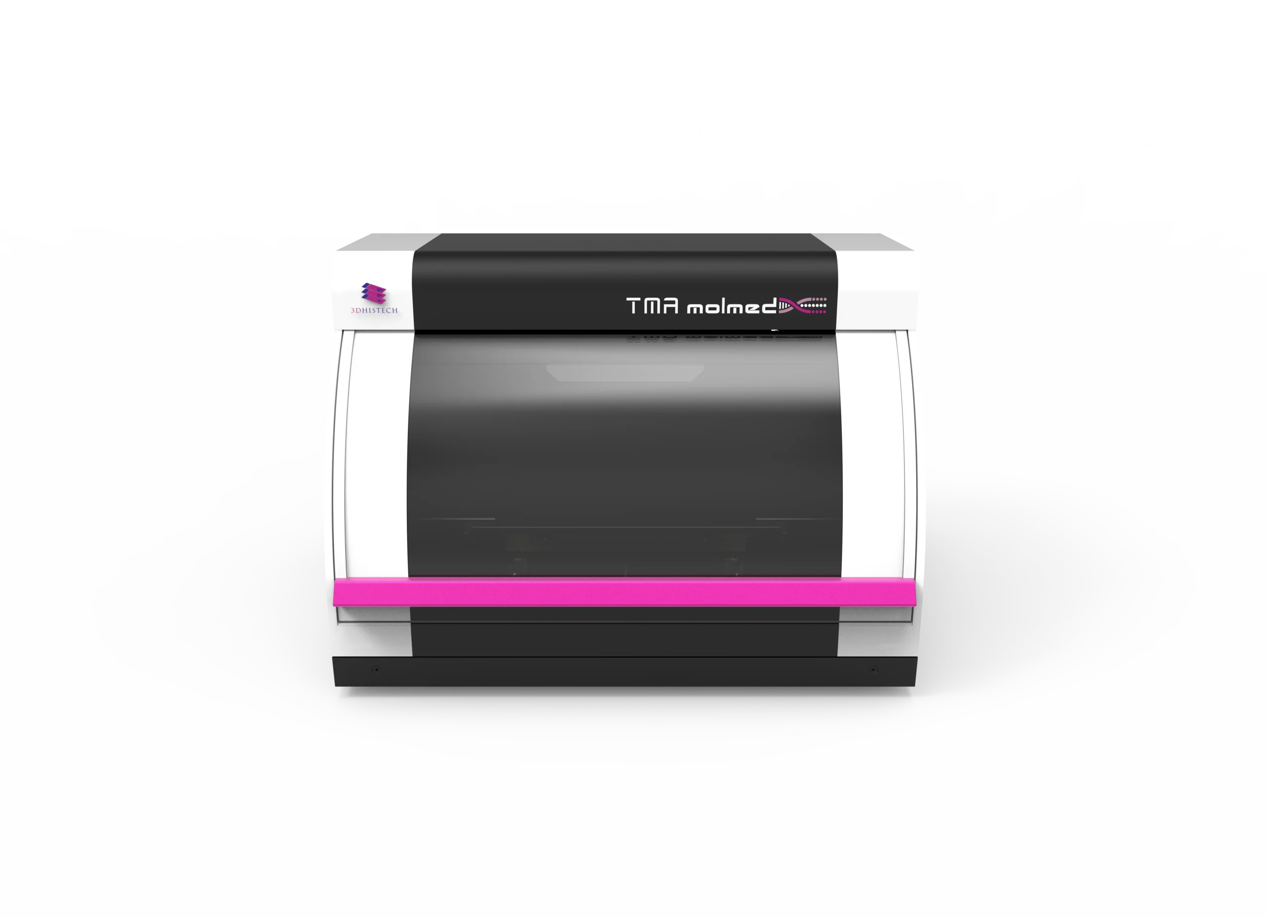
TMA molmed – Modular Precision for Tissue Microarray Creation & Molecular Sampling

The TMA molmed is a fully automated, high-throughput system that seamlessly integrates tissue microarray (TMA) creation with laser microdissection-like molecular sampling, making it a powerful tool for molecular pathology, translational research, and biobanking. Its modular architecture accommodates six core sizes as small as 0.2 mm, offering unmatched flexibility for laboratories handling both standard and mega blocks.

With mega block compatibility, this dual-functionality instrument enables high-volume sample processing, supporting up to six standard donor blocks or two mega blocks with 30 PCR tubes, as well as configurations for three standard donor blocks or one mega block with a wellplate. Its open modular architecture supports PCR cassettes, microtiter plates, and customized tissue cassettes, ensuring seamless integration into molecular workflows for DNA, RNA, and protein extraction, including PCR, dPCR, and sequencing applications.

Engineered for efficiency and precision, the TMA molmed transfers 200-250 cores per hour and supports high-density TMA block creation, accommodating up to 558 cores per recipient block. Automated block height measurement ensures proper core alignment, while digital integration with MRXS digital slides and JPEG overlays enhances accuracy in tissue selection. A comprehensive data management system streamlines workflows with barcode tracking, automated project data saving, and multi-format export options.

Combining modular adaptability with high-speed automation, the TMA molmed sets new standards in TMA preparation and molecular research. Its customizable recipient block designs, six configurable core sizes, and automated molecular sampling to PCR tubes or wellplates ensure unparalleled precision, efficiency, and contamination-free sample handling for state-of-the-art laboratory applications.

Key Features:

Dual-Functionality for Tissue Microarraying and Molecular Sampling
  • Combines high-precision tissue microarray creation with laser microdissection-like molecular sampling
  • Supports both standard and mega blocks, ensuring flexibility for various research applications
  • Six configurable core sizes (as small as 0.2 mm) for precise sample extraction
  • Mega block compatibility allows high-volume processing
  • Up to 6 standard donor blocks or 2 mega blocks with 30 PCR tubes
  • Configurations for 3 standard donor blocks or 1 mega block with a wellplate
  • Open modular design supports PCR cassettes, microtiter plates, and customized tissue cassettes


  • Transfers 200-250 cores per hour, optimizing workflow efficiency
  • Accommodates up to 558 cores per recipient block, supporting high-density TMAs
  • Automated block height measurement ensures precise alignment of tissue cores
  • MRXS digital slide and JPEG overlay compatibility for accurate tissue core selection
  • Barcode tracking, automated donor block imaging, and data saving enhance workflow efficiency
  • TMA Control Software enables custom recipient block design and real-time project tracking
  • Direct molecular sample extraction to PCR tubes or wellplates, eliminating cross-contamination risks
  • Supports DNA, RNA, and protein extraction, enabling downstream applications like PCR, dPCR, and sequencing
  • Multi-format data export (ods, xls, xlsx, csv, xml) for seamless integration into laboratory information systems
Capacity
  • TMA Blocks (Donor or Recipient): 9
  • Macro Donor or Recipient Blocks: 2
  • TMA Mega Donor Blocks: 6
  • PCR Tube Capacity: 30
  • Wellplate Capacity: 1 wellplate
Core Size & Density
  • Core Diameters (mm): 0.2 (surface), 0.6, 1.0, 1.5, 2.0, 3.0
  • Maximum Number of Cores per TMA Block:
  • 6 mm: 558 cores
  • mm: 286 cores
  • 5 mm: 135 cores
  • 0 mm: 84 cores
  • 0 mm: 40 cores
Performance

Speed: 200-250 cores per hour

Barcode & Data Handling
  • Barcode Reading: 1D and 2D (optional)
  • Supported 1D Codes: Code 39, UPC-A, EAN 13, Codabar, Code 128, etc.
  • Supported 2D Codes: Data Matrix, PDF417, QR Code, Aztec, MicroQR, etc.
  • Supported Import Formats: .XLS, .XLSX, .ODS
  • Supported Export Formats: .XLS, .XLSX, .ODS, .CSV, .XML
Dimensions & Weight
  • Base Unit Dimensions (W x D x H): 552 x 400 x 400 mm
  • Toolbox Dimensions: 310 x 110 x 155 mm
  • Power Supply Unit Dimensions: 189 x 90 x 44 mm
  • Base Unit Weight: 29 kg
  • Toolbox Weight: 5 kg
  • Display Weight: 7.1 kg
  • Power Supply Unit Weight: 0.55 kg
Camera & Image Digitization
  • Camera: 12 MP Blackfly S USB3 digital imaging camera (Sony IMX226)
  • Resolution: 4000 x 3000 pixels
  • Pixel Size: 1.85 µm
  • Frame Rate: 31 FPS
  • Connection: USB 3.0
Light Source
  • Type: Multichannel RGB LED light source (transmitted light operation)
Computer System Requirements
  • Processor: Intel Core i9-14900 (24 cores, 32 threads) or similar
  • RAM: 64 GB
  • SSD Storage: 256 GB
  • HDD Storage: 2 TB
  • Graphics Output: 2x DisplayPort (4096 x 2304)
  • Ports: 4x USB 3.2, 2x USB 2.0, 1x Ethernet (10/100/1000)
  • Operating System: Windows 11 Pro (64-bit)
Accessories
  • Base Molds for Paraffin Blocks: 3 pcs
  • Calibration Blocks (Normal/Mega): 1/1 pcs
  • Modules:
  • Drill Module (Takes 3 slots)
  • Normal Module (Takes 1 slot)
  • Wellplate Module (Takes 2 slots)
  • Mega Module (Takes 1 slot)
  • PCR Module (Takes 1 slot)
  • Punch Needles (mm): 0.2, 0.6, 1.0, 1.5, 2.0, 3.0 (3 pcs each)
  • Punch Pistons (mm): 0.2, 0.6, 1.0, 1.5, 2.0, 3.0 (3 pcs each)
Software
  • TMA Control Software – Manages TMA block design & creation
  • SlideViewer – Digital Microscopy Software
  • SlideMaster – Digital Slide Converter
  • Features:
  • Layout design & tissue core selection
  • Donor slide overlay with TMA markers
  • Automated slide search & overlay
  • Manual overlay option
  • Barcode integration
TMA molmed

Specifications

Use Cases of TMA molmed

Explore the Use Cases

Archaeology and paleontology have traditionally relied on destructive sampling methods such as sectioning, staining, and thin-section petrography to study fossils and artifacts at the microscopic level. However, these approaches pose risks to…
Biomedical research is experiencing a major shift with the integration of Digital Pathology and Micro-CT solutions, significantly improving tissue analysis, biomarker discovery, and disease modeling. Traditional microscopy methods have inherent limitations, such…
Rapid and accurate biopsy diagnostics and frozen section evaluations are critical for clinical decision-making, especially in oncology and intraoperative pathology. 3DHISTECH’s Pannoramic DX digital pathology solutions provide a complete workflow for digital…
Botany and plant sciences rely on high-resolution imaging and precise analysis of plant tissues to understand disease mechanisms, stress responses, genetic modifications, and ecological changes. Traditional microscopy techniques limit research efficiency due…
Cancer research demands cutting-edge imaging technologies to enhance biomarker discovery, tumor profiling, and immuno-oncology advancements. Traditional histopathology techniques, while fundamental, present challenges such as limited scalability, subjective interpretation, and data fragmentation. Digital…
As pathology departments face increasing case volumes and demands for faster diagnoses, traditional workflows can struggle with time-intensive slide analysis, inconsistent imaging, and limited remote collaboration. Digital Pathology offers a modern solution,…
Cytopathology plays a critical role in diagnosing cancer, infectious diseases, and other cellular abnormalities. The transition from conventional slide review to digital pathology enhances efficiency, precision, and collaboration in cytological diagnostics….
Developmental biology is a rapidly evolving field that focuses on understanding how organisms grow, develop, and differentiate at the cellular and molecular levels. Researchers face key challenges such as capturing high-resolution images…
Immunohistochemistry (IHC) plays a critical role in cancer diagnostics, biomarker discovery, and personalized medicine, requiring high-precision imaging, consistent staining interpretation, and workflow efficiency. 3DHISTECH’s Pannoramic DX Whole Slide Imaging (WSI) solutions provide…
Toxicological pathology and neuropathology require high-precision, high-throughput imaging solutions to assess tissue morphology, detect subtle abnormalities, and streamline data-intensive workflows. 3DHISTECH’s Pannoramic DX Digital Scanners provide fully automated, highresolution whole slide imaging…
Accurate diagnosis and efficient workflow are critical in gastrointestinal (GI) and liver pathology, where identifying subtle tissue changes can determine patient outcomes. The Pannoramic DX Digital Pathology Solutions featuring the Pannoramic 250…
Genetic research demands precise imaging tools capable of capturing high-resolution cellular and molecular structures for in-depth analysis. 3DHISTECH’s Solutions for digital pathology supported by QuantCenter, QuantServer, SlideManager, and SlideCenter, provide an AI-driven,…
Geological and mineralogical research relies on high-resolution imaging techniques to analyze mineral composition, rock microstructures, and sedimentary formations. Traditional petrographic microscopy, while effective, is often limited by sample preparation constraints, depth resolution,…
Immunology and infectious disease research require precise cellular analysis, biomarker quantification, and advanced multiplex imaging to understand immune responses, pathogen-host interactions, and disease progression. Traditional histopathology methods are labor-intensive, slow, and prone…
Molecular cell biology research relies on high-resolution imaging and fluorescence capabilities to analyze cellular structures, molecular markers, and complex biological interactions. The integration of digital pathology solutions from 3DHISTECH—Pannoramic Confocal, Pannoramic 250…
Neuroscience research demands high-resolution imaging, precise molecular analysis, and largescale data management to study the complexity of the brain, nervous system, and neurological diseases. Traditional microscopy and pathology techniques often limit the…
The fight against cancer demands precision, efficiency, and collaboration in pathology. The Pannoramic DX Whole Slide Imaging Solutions, including the Pannoramic 250 Flash III DX, Pannoramic 150 DX, and Pannoramic 75 DX…
Pharmacology research is essential for drug discovery, preclinical evaluation, and biomarker validation. However, researchers face significant challenges in high-throughput tissue analysis, biomarker quantification, and data integration. Traditional microscopy and manual pathology workflows…
Advancing telepathology and remote consultation, the Pannoramic DX Digital Pathology Solutions provide an end-to-end digital pathology workflow, ensuring rapid, high-resolution scanning and seamless remote collaboration. These solutions support real-time diagnostics, expert consultations,…
Toxicology research is essential for understanding the effects of drugs, chemicals, and environmental pollutants on biological systems. Traditionally, toxicology studies rely on manual histological evaluation, which can be time-consuming, prone to subjectivity,…
Toxicology and environmental science research requires high-precision imaging, high-throughput analysis, and advanced molecular assessments to evaluate the impact of toxins, chemicals, and environmental pollutants on biological systems. Traditional microscopy and histopathology methods…
Veterinary pathology plays a crucial role in diagnosing diseases in animals, supporting clinical treatment, biomedical research, and toxicologic pathology. The Pannoramic DX Digital Pathology solutions from 3DHISTECH provide high-speed, high-resolution digital pathology…
Veterinary pathology plays a vital role in animal disease research, wildlife conservation, livestock farming, and food safety. As disease outbreaks, environmental changes, and food security challenges rise, digital pathology, AI-driven analysis, and…

Still thinking?

Ask us a question about TMA molmed

Ask us a question

Request Demo