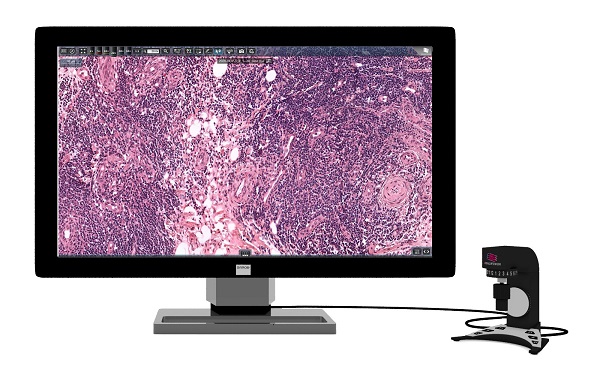
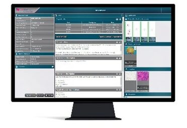
Diagnostic Applications is an image analysis platform designed as a decision support tool for fast, field of view-based, automated quantification.
With the help of this computer-aided image analysis tool, accurate, fast, and high-quality analytical results can be generated. Thanks to the integration of CaseManager and Diagnostic Applications, the measurement results, charts and masked images of the measurement area are automatically displayed in the appropriate pathology case on CaseManager’s interface. The pathologist can easily attach the measurement results and images to the final report.
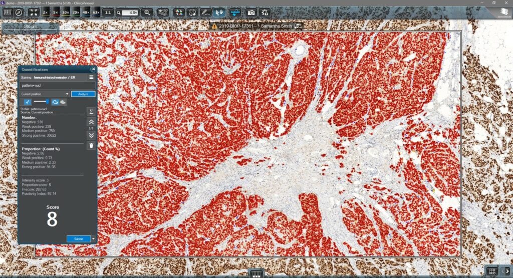
The image analysis modules integrated into ClinicalViewer provide the pathologist with a quantitative image analysis framework: Four modules (Estrogen, HER2, Ki67* and Progesterone) help quantify solid tumor cases. The results of the analysis are then included in the final report in CaseManager.
Key features
- Four image analysis tools (Er, Pr, HER2, Ki67*) offer image analysis solutions designed for diagnostic breast panel assessment with the IVD-approved MembraneQuant and NuclearQuant algorithms inside
- Express image analysis process on current view or predefined annotation
- Measurement result saving process: the result of the image evaluation is saved to the patient data
Further available image analysis tools
PatternQuant
Pattern-based machine learning algorithm that can be trained with region of interest (ROI) definition for both brightfield and fluorescent tissue classification or pre-segmentation tasks.
CellQuant
Pattern-based machine learning algorithm that can be trained with region of interest (ROI) definition for both brightfield and fluorescent tissue classification or pre-segmentation tasks.
HistoQuant
Segmentation algorithm for cells/membrane/cytoplasm or whole cell, based on color deconvolution; optional classification based on chromogen quantity in any of the segmented cell compartments.
*For performance evaluation
